Advanced Ultrasound Technology at Orbit Diagnostics & Healthcare
At Orbit Diagnostics & Healthcare, we believe that accurate diagnosis begins with high-quality imaging. That is why we have invested in the GE Healthcare Versana Balance ultrasound system, a modern diagnostic platform designed to deliver exceptional image clarity, advanced Doppler capabilities and versatile clinical applications. Our commitment is simple: provide patients and referring doctors with reliable diagnostic information supported by modern medical technology and professional expertise.
Why We Invest in Advanced Ultrasound Technology
When patients undergo an ultrasound examination, most only see the final images and report. Behind every image, however, is a sophisticated combination of hardware, software, signal processing and imaging technologies working together to create the diagnostic information used by healthcare professionals.
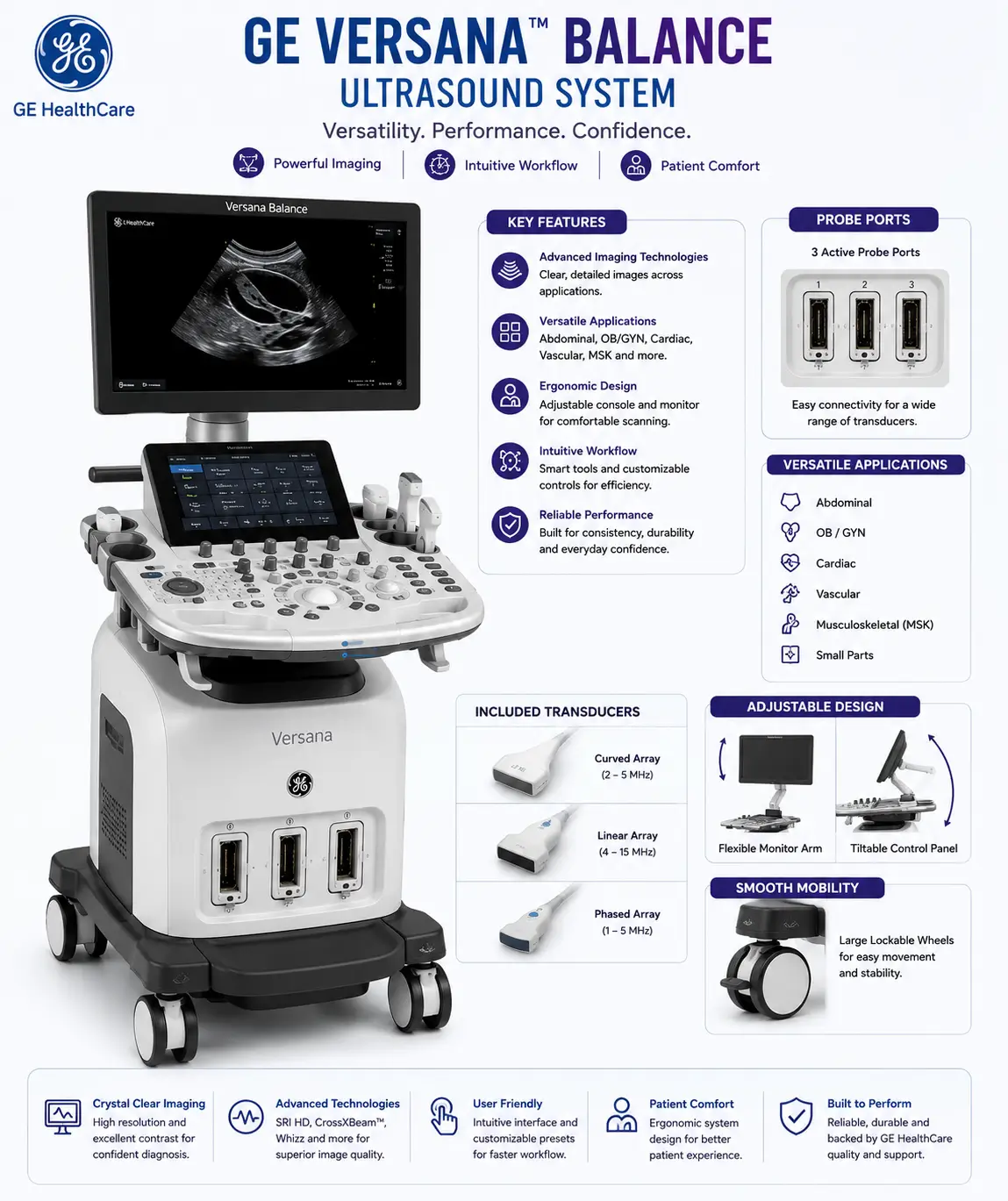
Modern ultrasound systems have evolved significantly over the years. Improvements in image processing, Doppler technology, probe design and digital optimisation now allow healthcare professionals to visualise anatomical structures with greater detail and consistency than ever before.
At Orbit Diagnostics & Healthcare, we selected the GE Versana Balance because it offers a powerful combination of image quality, clinical versatility and workflow efficiency. Whether a patient is visiting us for a pregnancy ultrasound, thyroid scan, abdominal examination, vascular Doppler study, transvaginal ultrasound or echocardiography assessment, our objective remains the same: provide reliable imaging that supports accurate diagnostic evaluation.
Technology alone cannot make a diagnosis. However, when advanced imaging equipment is combined with experienced healthcare professionals and careful examination techniques, it creates a strong foundation for high-quality diagnostic care.
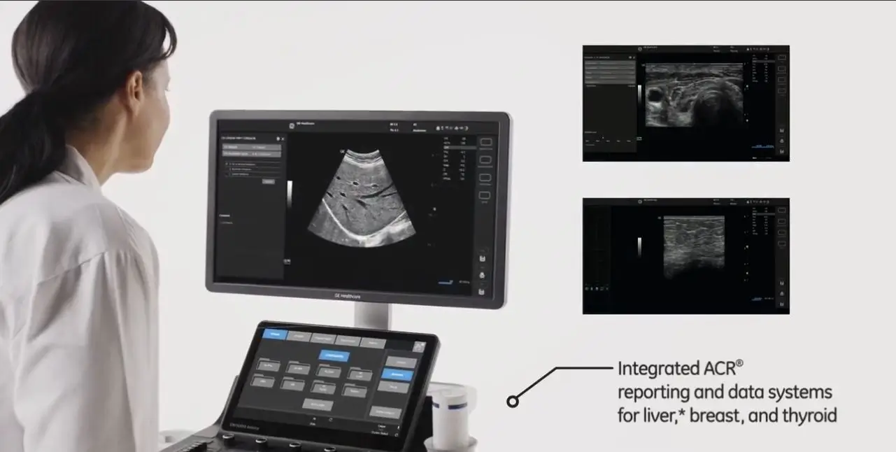
Why Ultrasound Technology Matters
Not all ultrasound systems are the same. The quality of the imaging technology being used can influence how clearly anatomical structures are visualised and how efficiently examinations are performed. Clear imaging helps healthcare professionals evaluate organs, blood vessels, soft tissues and pregnancy-related structures with greater confidence.
Think of it like photography. A blurry image may provide some information, but a sharper image often reveals important details that would otherwise be difficult to appreciate. Ultrasound imaging follows a similar principle. Improved image clarity can help support more detailed assessment and accurate documentation of findings.
For patients, this means examinations performed using technology designed to provide detailed visualisation across a broad range of clinical applications. For healthcare professionals, it means access to imaging tools that help support confident interpretation and reporting.
Designed to provide detailed visualisation of organs, tissues and anatomical structures.
Supports blood flow assessment in vascular, pregnancy and cardiac studies.
Dedicated probes for abdominal, thyroid, vascular, pregnancy and cardiac imaging.
Touchscreen controls and intelligent optimisation tools improve efficiency.
The GE Versana Balance Ultrasound Platform
At Orbit Diagnostics & Healthcare, we use the GE Healthcare Versana Balance ultrasound system because it combines modern imaging technology, workflow efficiency and diagnostic versatility within a single platform. Designed by GE Healthcare, one of the world's leading medical imaging manufacturers, the Versana Balance has been developed to support a wide range of clinical applications while maintaining consistent image quality and dependable performance.
Rather than being designed for only one speciality, the system supports abdominal imaging, pregnancy ultrasound, thyroid examinations, breast imaging, vascular Doppler studies, musculoskeletal assessments, pelvic ultrasound and cardiac imaging. This flexibility allows our team to perform diverse examinations while maintaining a consistent standard of diagnostic quality.
For patients, this means access to modern ultrasound technology capable of supporting a broad range of healthcare needs. For healthcare professionals, it provides the tools required to perform examinations efficiently while maintaining confidence in image quality and reporting.
Modern Touchscreen Technology for Faster Workflow
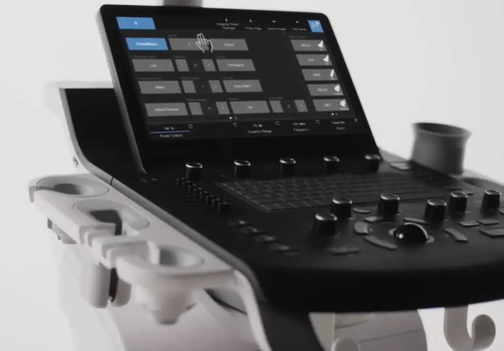
One of the most noticeable features of the GE Versana Balance is its modern touchscreen control system. Traditional ultrasound machines often relied heavily on complex button layouts and multiple control panels. The Versana Balance introduces a more streamlined approach, allowing healthcare professionals to access commonly used functions quickly and efficiently.
Measurements, imaging presets, patient information, Doppler settings and examination tools can be accessed through an intuitive interface designed to reduce unnecessary workflow interruptions. This allows the examiner to spend more time focusing on image acquisition and patient care rather than navigating complicated system controls.
The practical benefit for patients is often a smoother examination experience with efficient image capture and reduced delays during scanning.
Compact Design with Clinical Mobility
Healthcare environments require flexibility. The GE Versana Balance has been engineered with a compact footprint that allows it to fit comfortably within modern clinical settings while maintaining powerful imaging capabilities.
Despite its streamlined design, the system incorporates advanced imaging technologies that would traditionally have been found only in much larger platforms. This combination of performance and mobility allows examinations to be performed efficiently without sacrificing image quality.
| Feature | Clinical Advantage |
|---|---|
| Compact Footprint | Efficient use of examination room space |
| Mobile Platform | Flexible positioning during examinations |
| Integrated Workflow Design | Improved scanning efficiency |
| Multi-Probe Support | Versatile clinical applications |
| Modern Ergonomics | Enhanced operator comfort during prolonged examinations |
These design considerations help create a more comfortable environment for both patients and healthcare professionals.
High-Resolution Imaging Designed for Diagnostic Confidence
The true value of an ultrasound system lies in the quality of the images it produces. During an ultrasound examination, healthcare professionals often evaluate very small anatomical details. The ability to clearly visualise these structures can play an important role in clinical assessment.
The GE Versana Balance incorporates advanced digital imaging technologies designed to improve image sharpness, tissue differentiation and anatomical visualisation. These technologies help create images that are detailed, consistent and clinically useful across a wide range of examinations.
Whether evaluating the liver, kidneys, thyroid gland, blood vessels, musculoskeletal structures or a developing baby during pregnancy, clear imaging supports careful examination and accurate documentation of findings.
Improved differentiation between adjacent anatomical structures.
Reliable visualisation across different examination types.
Better distinction between tissues and anatomical boundaries.
Supports visualisation of structures located deeper within the body.
Powerful Digital Processing Working Behind Every Image
Modern ultrasound imaging is far more sophisticated than simply transmitting sound waves into the body. Every ultrasound image undergoes complex processing before it appears on the monitor.
The GE Versana Balance incorporates advanced digital signal processing technologies that analyse incoming ultrasound information in real time. These systems continuously optimise image presentation, helping healthcare professionals visualise anatomy more effectively.
Beamforming technology, digital image optimisation and advanced processing algorithms work together to improve image consistency and clarity. The result is imaging that helps support confident interpretation and efficient reporting.
Although patients may never see these technologies directly, they play an important role in creating the high-quality diagnostic images used during everyday clinical practice.
Why Healthcare Professionals Appreciate the GE Versana Balance
Diagnostic imaging professionals spend countless hours performing and interpreting ultrasound examinations. The quality of the equipment they use every day has a direct impact on workflow, efficiency and image acquisition.
The GE Versana Balance has been designed to help simplify routine tasks while providing access to advanced imaging capabilities. Features such as intelligent workflow tools, responsive controls, structured examination presets and streamlined reporting support efficient clinical practice.
When technology becomes easier to use, healthcare professionals can focus more attention on image interpretation, patient communication and diagnostic decision-making.
This balance between technological sophistication and practical usability is one of the reasons the GE Versana Balance has become a trusted ultrasound platform in many healthcare environments around the world.
Specialised Ultrasound Probes for Different Diagnostic Applications
One of the major advantages of the GE Healthcare Versana Balance is its ability to support multiple specialised ultrasound probes. Different organs, tissues and clinical applications require different imaging frequencies and scanning techniques. A single probe cannot effectively perform every type of ultrasound examination.
To ensure optimal image quality across a wide range of clinical applications, Orbit Diagnostics & Healthcare uses dedicated probes designed for specific anatomical regions and examination types. This allows healthcare professionals to select the most appropriate transducer for each patient, helping achieve detailed visualisation and reliable diagnostic information.
| Probe | Primary Applications | Strength |
|---|---|---|
| Convex Probe | Abdominal, Obstetric, Pelvic Ultrasound | Deep penetration and wide field of view |
| Linear Probe | Thyroid, Breast, Doppler, Soft Tissue | High-resolution superficial imaging |
| TVS Probe | Transvaginal Ultrasound and Fertility Studies | Detailed pelvic visualisation |
| Cardiac Probe | Echocardiography and Cardiac Assessment | Optimised heart imaging |
Convex Probe: The Foundation of Abdominal and Pregnancy Ultrasound
The convex probe is one of the most frequently used ultrasound transducers in modern diagnostic imaging. Its curved design enables the ultrasound beam to cover a wide area while providing excellent penetration into deeper structures within the body.
This makes it particularly valuable for evaluating abdominal organs and monitoring pregnancy. The probe can visualise structures located deeper within the abdomen while maintaining reliable image quality across a broad field of examination.
For many patients, this is the probe used during routine abdominal scans, pregnancy ultrasounds and pelvic evaluations.
| Scans Performed Using Convex Probe |
|---|
| Whole Abdomen Ultrasound |
| Upper Abdomen Ultrasound |
| Lower Abdomen Ultrasound |
| Liver Ultrasound |
| Gallbladder Ultrasound |
| Kidney Ultrasound |
| Urinary Bladder Ultrasound |
| Prostate Ultrasound |
| Pregnancy Ultrasound |
| NT Scan |
| Level II Anomaly Scan |
| Growth Scan |
| Biophysical Profile |
| Pelvic Ultrasound |
Linear Probe: Exceptional Detail for Thyroid, Breast and Doppler Studies
The linear probe operates at higher ultrasound frequencies than a convex probe. While higher frequencies do not penetrate as deeply into the body, they produce remarkably detailed images of structures located near the skin surface.
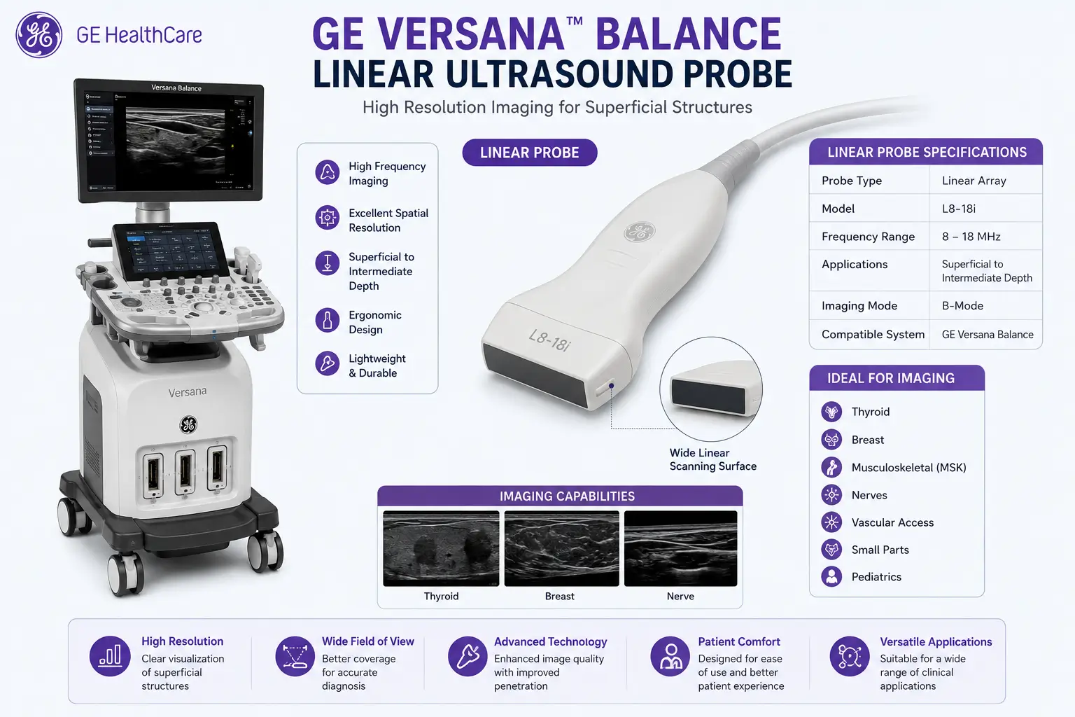
This makes the linear probe ideal for examinations where fine anatomical detail is important. The probe is particularly valuable for evaluating the thyroid gland, breast tissue, lymph nodes, tendons, muscles and blood vessels.
Its ability to provide excellent spatial resolution helps healthcare professionals assess small structures with greater confidence.
| Scans Performed Using Linear Probe |
|---|
| Thyroid Ultrasound |
| Breast Ultrasound |
| Neck Ultrasound |
| Lymph Node Assessment |
| Soft Tissue Ultrasound |
| Musculoskeletal Ultrasound |
| Tendon Evaluation |
| Joint Assessment |
| Carotid Doppler Study |
| Peripheral Arterial Doppler |
| Peripheral Venous Doppler |
| Varicose Vein Assessment |
| Scrotal Ultrasound |
TVS Probe: High-Resolution Women's Health and Fertility Imaging
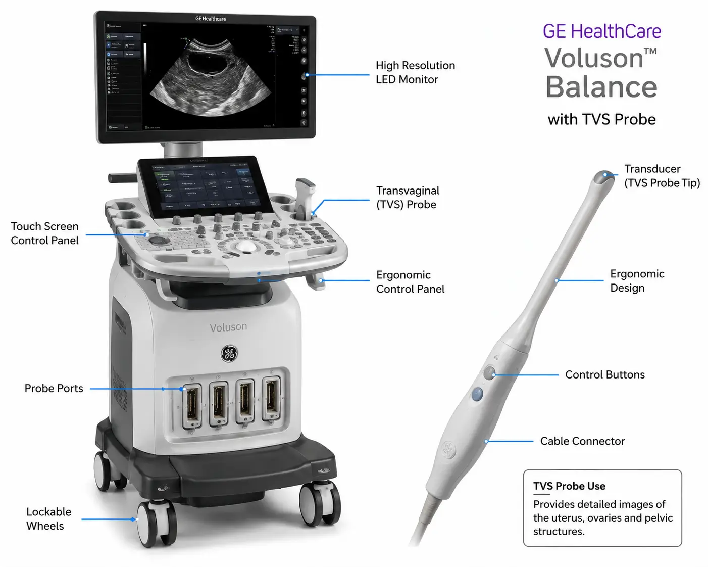
The Transvaginal Ultrasound (TVS) probe allows the ultrasound transducer to be positioned closer to the pelvic organs being evaluated. This often results in significantly improved image detail compared to conventional abdominal pelvic ultrasound examinations.
The probe is widely used in gynaecology, infertility evaluation and early pregnancy assessment where high-resolution pelvic imaging is essential.
| Scans Performed Using TVS Probe |
|---|
| Transvaginal Ultrasound (TVS) |
| Early Pregnancy Scan |
| Viability Scan |
| Follicular Monitoring |
| Infertility Assessment |
| Ovarian Evaluation |
| Endometrial Assessment |
| Uterine Evaluation |
| PCOS Assessment |
| Pelvic Pain Evaluation |
Cardiac Probe: Dedicated Technology for Echocardiography
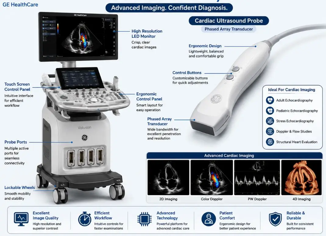
The heart is a constantly moving organ that requires specialised imaging techniques. Standard probes are not optimised for detailed cardiac imaging, which is why dedicated cardiac transducers are used for echocardiography.
The cardiac probe utilised with the GE Versana Balance has been designed to visualise cardiac chambers, heart valves, blood flow and cardiac function while maintaining excellent temporal resolution.
This specialised technology enables comprehensive cardiac assessment and supports a wide range of echocardiographic applications.
| Cardiac Examinations Performed |
|---|
| 2D Echocardiography |
| Heart Chamber Assessment |
| Valve Evaluation |
| Cardiac Function Assessment |
| Colour Flow Imaging |
| Cardiac Doppler Studies |
| Ejection Fraction Assessment |
| Pericardial Evaluation |
The Right Probe for the Right Examination
A high-quality ultrasound machine is only part of the equation. Selecting the appropriate probe for each examination is equally important. Different anatomical structures require different frequencies, depths of penetration and imaging characteristics.
At Orbit Diagnostics & Healthcare, every ultrasound examination is performed using the most appropriate probe for the clinical indication. This approach helps optimise image quality and ensures that the imaging technology is being used to its full potential.
Combined with the advanced imaging capabilities of the GE Versana Balance, our specialised probe selection allows us to support a broad range of diagnostic ultrasound services while maintaining a strong focus on image quality and diagnostic confidence.
The Technology Working Behind Every Ultrasound Image
When patients see an ultrasound image on the screen, they are looking at the final result of thousands of calculations taking place every second. Modern ultrasound systems do much more than simply send sound waves into the body and display a picture.
The GE Healthcare Versana Balance combines advanced signal processing, image optimisation technologies and intelligent workflow tools designed to improve image clarity and support diagnostic confidence. These technologies continuously analyse incoming ultrasound information and help create images that are clearer, more detailed and more clinically useful.
At Orbit Diagnostics & Healthcare, these capabilities help us provide high-quality imaging for pregnancy ultrasound, abdominal examinations, thyroid imaging, vascular Doppler studies, cardiac assessments and many other applications.
Tissue Harmonic Imaging (THI)
Tissue Harmonic Imaging, commonly known as THI, is one of the most important advancements in modern ultrasound imaging. Rather than relying solely on conventional ultrasound signals, the system processes harmonic frequencies generated naturally within body tissues.
The result is often improved image quality with reduced noise and better visualisation of anatomical structures. This can be particularly valuable when evaluating deeper organs or imaging patients where standard ultrasound techniques may be more challenging.
| Benefits of THI |
|---|
| Improved image clarity |
| Reduced artefacts |
| Better tissue differentiation |
| Enhanced contrast resolution |
| Improved visualisation of deeper structures |
CrossXBeam™ Technology
CrossXBeam™ is GE Healthcare's spatial compounding technology. Instead of creating an image from a single beam direction, the system combines information from multiple ultrasound beam angles.
This process helps reduce image artefacts while improving tissue border definition and anatomical detail. Structures often appear more clearly outlined, helping healthcare professionals evaluate anatomy with greater confidence.
| Conventional Imaging | CrossXBeam™ Imaging |
|---|---|
| Single beam angle | Multiple beam angles combined |
| More image artefacts | Reduced artefacts |
| Less tissue definition | Improved tissue borders |
| Variable image consistency | More consistent visualisation |
For patients, the technology works quietly in the background. For healthcare professionals, it can help reveal anatomical details that might otherwise be more difficult to appreciate.
Speckle Reduction Imaging (SRI)
Ultrasound images naturally contain a granular appearance known as speckle. While some speckle is unavoidable, excessive speckle can sometimes make it harder to visualise subtle anatomical details.
Speckle Reduction Imaging technology analyses image data and reduces unnecessary noise while preserving important diagnostic information. This helps produce cleaner images with improved visual clarity.
Sharper tissue boundaries and improved organ visualisation can contribute to more confident image interpretation and reporting.
Reduced visual noise during imaging.
Better visualisation of anatomical structures.
Improved edge clarity.
Supports more detailed assessment.
Advanced Color Doppler Technology
Understanding blood flow is an essential component of many ultrasound examinations. The GE Versana Balance includes advanced Color Doppler technology that allows healthcare professionals to visualise blood circulation in real time.
Different colours displayed on the screen help represent blood movement and flow direction, providing additional information beyond standard grayscale imaging.
This technology plays an important role in vascular studies, thyroid imaging, pregnancy assessments, abdominal examinations and cardiac evaluations.
| Clinical Application | How Color Doppler Helps |
|---|---|
| Pregnancy Ultrasound | Assessment of placental and fetal circulation |
| Vascular Studies | Evaluation of blood vessels and blood flow |
| Thyroid Imaging | Assessment of vascular patterns |
| Abdominal Imaging | Evaluation of organ blood supply |
| Cardiac Imaging | Visualisation of blood flow through the heart |
Power Doppler Technology
Some blood vessels contain slower or more subtle blood flow signals that may be difficult to visualise using conventional techniques. Power Doppler technology is designed to improve sensitivity to these blood flow signals.
By increasing sensitivity to vascular information, the system can help visualise smaller vessels and lower-flow circulatory patterns that may be clinically relevant during certain examinations.
This additional capability expands the diagnostic versatility of the ultrasound platform and supports more comprehensive vascular assessment.
Pulsed Wave and Spectral Doppler Analysis
While Color Doppler provides a visual representation of blood flow, Pulsed Wave Doppler allows healthcare professionals to measure and analyse blood flow characteristics at specific locations.
The technology provides detailed information regarding blood flow velocity and waveform patterns, helping support vascular and cardiac assessments.
This quantitative information complements conventional imaging and contributes to a more comprehensive understanding of circulatory dynamics.
Intelligent Image Optimisation Tools
Achieving optimal ultrasound images often requires adjustments to brightness, gain, contrast and other imaging parameters. The GE Versana Balance includes intelligent optimisation tools that assist in obtaining high-quality images efficiently.
These technologies automatically analyse image characteristics and help optimise settings for improved visualisation. This can help reduce workflow complexity while maintaining image consistency across examinations.
For patients, this contributes to smoother examinations. For healthcare professionals, it supports efficiency and helps maintain imaging quality throughout the scanning process.
What These Technologies Mean for Patients
Most patients are not concerned about technical terminology or engineering specifications. What matters most is whether the technology being used can support a thorough and reliable examination.
The imaging technologies integrated into the GE Versana Balance are designed to improve image quality, enhance workflow efficiency and support detailed diagnostic evaluation. Combined with professional expertise and careful examination techniques, they help create an environment focused on quality patient care.
Improved visualisation of anatomical structures.
Advanced Doppler technologies support vascular evaluation.
Modern workflow tools help streamline scanning.
Technology designed to support reliable interpretation.
Why Image Quality Can Make a Meaningful Difference
Many people assume that all ultrasound machines produce similar images. In reality, there can be significant differences in image quality between older-generation systems and modern ultrasound platforms. While all ultrasound examinations rely on sound waves, the way those signals are processed, enhanced and displayed can have a substantial impact on the final image.
Modern imaging technology cannot replace clinical expertise, but it can provide healthcare professionals with clearer visual information. Better visualisation may help improve anatomical assessment, support accurate measurements and contribute to more confident diagnostic interpretation.
At Orbit Diagnostics & Healthcare, our investment in advanced ultrasound technology reflects our belief that patients deserve access to modern diagnostic tools capable of supporting high-quality imaging and reliable clinical evaluation.
How Better Imaging Helps in Everyday Clinical Practice
The benefits of advanced ultrasound technology become easier to appreciate when viewed through real clinical situations. Every day, healthcare professionals evaluate organs, tissues, blood vessels and developing babies using ultrasound imaging. In many of these situations, image clarity can play an important role in visualisation and assessment.
| Clinical Examination | How High-Quality Imaging Helps |
|---|---|
| Pregnancy Ultrasound | Improved visualisation of fetal anatomy, growth and pregnancy structures. |
| Level II Anomaly Scan | Detailed assessment of the fetal brain, heart, spine, kidneys and limbs. |
| Thyroid Ultrasound | Clearer visualisation of thyroid nodules and gland architecture. |
| Breast Ultrasound | Enhanced tissue visualisation and lesion assessment. |
| Liver Ultrasound | Better evaluation of liver texture and focal findings. |
| Kidney Ultrasound | Improved visualisation of stones, cysts and collecting systems. |
| Doppler Studies | More detailed assessment of blood flow characteristics. |
| Echocardiography | Clearer visualisation of cardiac chambers, valves and function. |
Although technology alone cannot determine a diagnosis, better visualisation helps healthcare professionals gather the information needed for careful evaluation and reporting.
The Advantage of High-Frequency Ultrasound Imaging
One of the key strengths of the GE Versana Balance system is its support for specialised high-frequency transducers. These probes are capable of producing highly detailed images of structures located near the skin surface, making them particularly valuable for thyroid imaging, breast ultrasound, vascular studies and soft tissue examinations.
High-frequency ultrasound provides exceptional spatial resolution, allowing healthcare professionals to evaluate smaller anatomical structures with remarkable detail. This level of visualisation can be particularly useful when assessing superficial organs and blood vessels where subtle findings may be clinically relevant.
Detailed assessment of thyroid tissue and nodules.
High-resolution visualisation of breast structures.
Detailed evaluation of arteries and veins.
Excellent visualisation of superficial tissues.
Older Ultrasound Systems vs Modern Ultrasound Technology
Medical imaging technology has advanced considerably over the years. Modern ultrasound platforms incorporate technologies that were not available in many earlier systems, helping improve image quality, workflow efficiency and overall examination capabilities.
| Feature | Older Generation Systems | GE Versana Balance |
|---|---|---|
| Image Processing | Earlier-generation technology | Advanced digital processing |
| Tissue Harmonic Imaging | Limited capability | Advanced implementation |
| Spatial Compounding | Often unavailable | CrossXBeam™ technology |
| Doppler Performance | Basic functionality | Advanced Doppler imaging |
| Workflow Tools | More manual adjustments | Intelligent optimisation features |
| Probe Versatility | Limited applications | Multiple specialised probes |
| Clinical Applications | More restricted | Broad multi-specialty support |
Why Orbit Diagnostics & Healthcare Invested in GE Healthcare Technology
Selecting diagnostic equipment is one of the most important decisions any healthcare provider makes. While many ultrasound systems are available in the market, our goal was not simply to purchase a machine. Our objective was to invest in technology capable of supporting high-quality diagnostic imaging across a wide range of clinical applications.
The GE Versana Balance was chosen because it combines advanced imaging capabilities, versatile probe support, intelligent workflow tools and the reliability associated with one of the world's most recognised healthcare technology manufacturers.
We wanted a platform capable of supporting routine examinations while also providing the imaging quality needed for specialised studies such as Level II Anomaly Scans, thyroid imaging, Doppler studies, transvaginal ultrasound examinations and echocardiography.
For us, investing in modern technology is part of a broader commitment to quality. It reflects our belief that patients deserve access to diagnostic equipment capable of supporting thorough evaluation and dependable reporting.
What This Means for You as a Patient
Most patients are not interested in technical specifications, engineering terminology or software architecture. What matters is whether the examination is being performed using reliable technology and whether the resulting information can assist their healthcare provider.
When you visit Orbit Diagnostics & Healthcare, your ultrasound examination is performed using a modern GE Healthcare platform designed to support a wide variety of diagnostic applications. Combined with specialised probes and advanced imaging technologies, the system helps provide detailed visualisation across pregnancy, abdominal, thyroid, vascular, breast, musculoskeletal and cardiac examinations.
More importantly, our commitment extends beyond technology itself. Every examination is performed with attention to detail, professional care and a focus on delivering accurate and clinically useful diagnostic information.
Advanced imaging technology trusted worldwide.
Designed for different organs and clinical applications.
Supports detailed blood flow assessment.
Technology combined with professional expertise.
Our Commitment to Advanced Diagnostic Technology
Healthcare technology continues to evolve, and Orbit Diagnostics & Healthcare remains committed to adopting solutions that support quality patient care. We continually evaluate technologies that can enhance imaging performance, workflow efficiency and diagnostic confidence.
The GE Versana Balance represents more than a piece of equipment. It reflects our commitment to providing diagnostic services supported by modern technology, professional expertise and a dedication to clinical excellence.
Whether you are visiting us for a routine abdominal ultrasound, a pregnancy scan, a thyroid examination, a Doppler study or an echocardiography assessment, you can be confident that your examination is being performed using technology selected with one objective in mind: helping provide reliable diagnostic information that supports informed healthcare decisions.
Frequently Asked Questions About Our Ultrasound Technology
Why does image quality matter in an ultrasound examination?
Image quality is one of the most important factors in diagnostic ultrasound. The clearer and more detailed the image, the easier it becomes for healthcare professionals to evaluate organs, tissues, blood vessels and developing babies. High-quality imaging helps improve visualisation of anatomical structures, supports accurate measurements and contributes to greater diagnostic confidence. While technology does not replace medical expertise, better image quality provides doctors with more reliable information for clinical interpretation and reporting.
Is GE Healthcare ultrasound technology considered reliable?
GE Healthcare is one of the world's most established medical technology companies and has been involved in diagnostic imaging for decades. Their ultrasound systems are widely used in hospitals, medical colleges, diagnostic centres and healthcare institutions across the world. The GE Versana Balance combines advanced imaging technologies, specialised clinical software and modern workflow tools designed to support high-quality diagnostic imaging across multiple medical specialties.
Is GE better than other ultrasound brands?
Several reputable ultrasound manufacturers produce excellent imaging systems, and each has its own strengths. What matters most is the combination of technology, image quality, clinical capabilities, maintenance support and the expertise of the healthcare professionals using the equipment. We selected the GE Versana Balance because it provides excellent image quality, advanced Doppler capabilities, versatile probe support and reliable performance across a broad range of clinical applications. Our focus is not on brand names alone but on providing technology that helps support accurate and dependable diagnostic evaluations.
What ultrasound examinations can be performed using the GE Versana Balance?
The GE Versana Balance is a versatile diagnostic ultrasound platform capable of supporting a wide variety of examinations. At Orbit Diagnostics & Healthcare, it is used for pregnancy ultrasound, Level II Anomaly Scan, Growth Scan, NT Scan, abdominal ultrasound, pelvic ultrasound, transvaginal ultrasound, thyroid ultrasound, breast ultrasound, soft tissue ultrasound, scrotal ultrasound, vascular Doppler studies, carotid Doppler, venous Doppler, arterial Doppler, musculoskeletal ultrasound, echocardiography and many other clinical applications.
Can the GE Versana Balance perform Color Doppler studies?
Yes. The GE Versana Balance includes advanced Color Doppler technology that allows healthcare professionals to visualise blood flow in real time. Color Doppler is commonly used during pregnancy assessments, thyroid evaluations, vascular studies, abdominal imaging and cardiac examinations. The system also supports additional Doppler technologies that provide valuable information regarding circulation and blood flow characteristics.
Can this machine perform Power Doppler examinations?
Yes. The system supports Power Doppler technology, which is designed to improve sensitivity to blood flow signals. This can be particularly useful when evaluating smaller blood vessels or lower-flow vascular structures that may require enhanced visualisation.
Can this machine perform cardiac ultrasound or echocardiography?
Yes. Using a dedicated cardiac probe, the GE Versana Balance can be used for echocardiography and cardiac assessments. These examinations may include evaluation of heart chambers, cardiac function, valve assessment, Doppler studies and other aspects of cardiac imaging. The specialised cardiac transducer has been designed specifically for heart imaging and allows detailed assessment of cardiac structures and blood flow.
Can the machine perform pregnancy ultrasounds?
Absolutely. The GE Versana Balance is well suited for obstetric imaging and supports a wide range of pregnancy ultrasound examinations, including Early Pregnancy Scans, Viability Scans, Dating Scans, NT Scans, Level II Anomaly Scans, Growth Scans and third-trimester assessments. Dedicated obstetric software helps support fetal measurements and pregnancy reporting workflows.
What probes are available at Orbit Diagnostics & Healthcare?
Our GE Versana Balance system is equipped with multiple specialised probes including a Convex Probe, Linear Probe, Transvaginal (TVS) Probe and Cardiac (Echo) Probe. Each probe is designed for specific clinical applications and helps ensure optimal image quality for different types of examinations.
Why did Orbit Diagnostics & Healthcare invest in this technology?
At Orbit Diagnostics & Healthcare, we believe diagnostic quality should never be compromised. We invested in the GE Versana Balance because it offers advanced imaging capabilities, modern workflow tools, specialised clinical software and excellent versatility across multiple ultrasound applications. Our goal is to provide patients and referring doctors with access to modern diagnostic imaging technology that supports accurate assessments and reliable reporting.
Can modern ultrasound technology improve diagnostic confidence?
Advanced imaging technologies can improve visualisation of anatomical structures, enhance image clarity and support more detailed examinations. While diagnosis ultimately depends on the expertise and judgement of healthcare professionals, better imaging can provide additional information that contributes to clinical confidence and decision-making.
Does better technology mean a better patient experience?
Modern ultrasound systems are designed not only to improve image quality but also to enhance workflow efficiency and patient comfort. Faster image optimisation, intuitive controls and specialised clinical software can contribute to smoother examinations while allowing healthcare professionals to focus more attention on patient care and diagnostic assessment.