Is The Anomaly Scan Painful?
No. The examination is generally painless. The ultrasound probe is moved across the abdomen while images are obtained using sound-wave technology.
Disclaimer: This page is intended to support patient understanding and should not replace professional medical advice, diagnosis or treatment.
For many expectant parents, the anomaly scan is one of the most anticipated appointments during pregnancy. It often arrives at a stage when the pregnancy feels more real, fetal movements may be becoming noticeable, and families are eager to learn more about their baby's development. Unlike earlier pregnancy ultrasounds that primarily confirm pregnancy and estimate gestational age, the anomaly scan is designed to provide a much more detailed assessment of fetal anatomy. During this examination, healthcare professionals carefully evaluate multiple parts of the baby's body, monitor growth and assess important structures that are developing during the second trimester. Many parents arrive with questions. What exactly happens during the scan? What are doctors looking for? How long does the examination take? Can every condition be detected? Understanding the purpose and process of the anomaly scan can help make the experience less stressful and far more meaningful.
Pregnancy involves several ultrasound examinations, each serving a different purpose. Early scans help confirm the pregnancy and establish gestational age. Later scans may be used to monitor growth and wellbeing. The anomaly scan occupies a unique position because it focuses on a detailed evaluation of the developing baby. During a single appointment, multiple organs and body structures can be assessed, providing valuable information about fetal development at that stage of pregnancy. For healthcare professionals, the examination provides important clinical information. For parents, it is often one of the first opportunities to see detailed images of their baby's face, limbs and movements. Because of its comprehensive nature, many healthcare providers consider the anomaly scan one of the most significant ultrasound examinations performed during routine prenatal care.
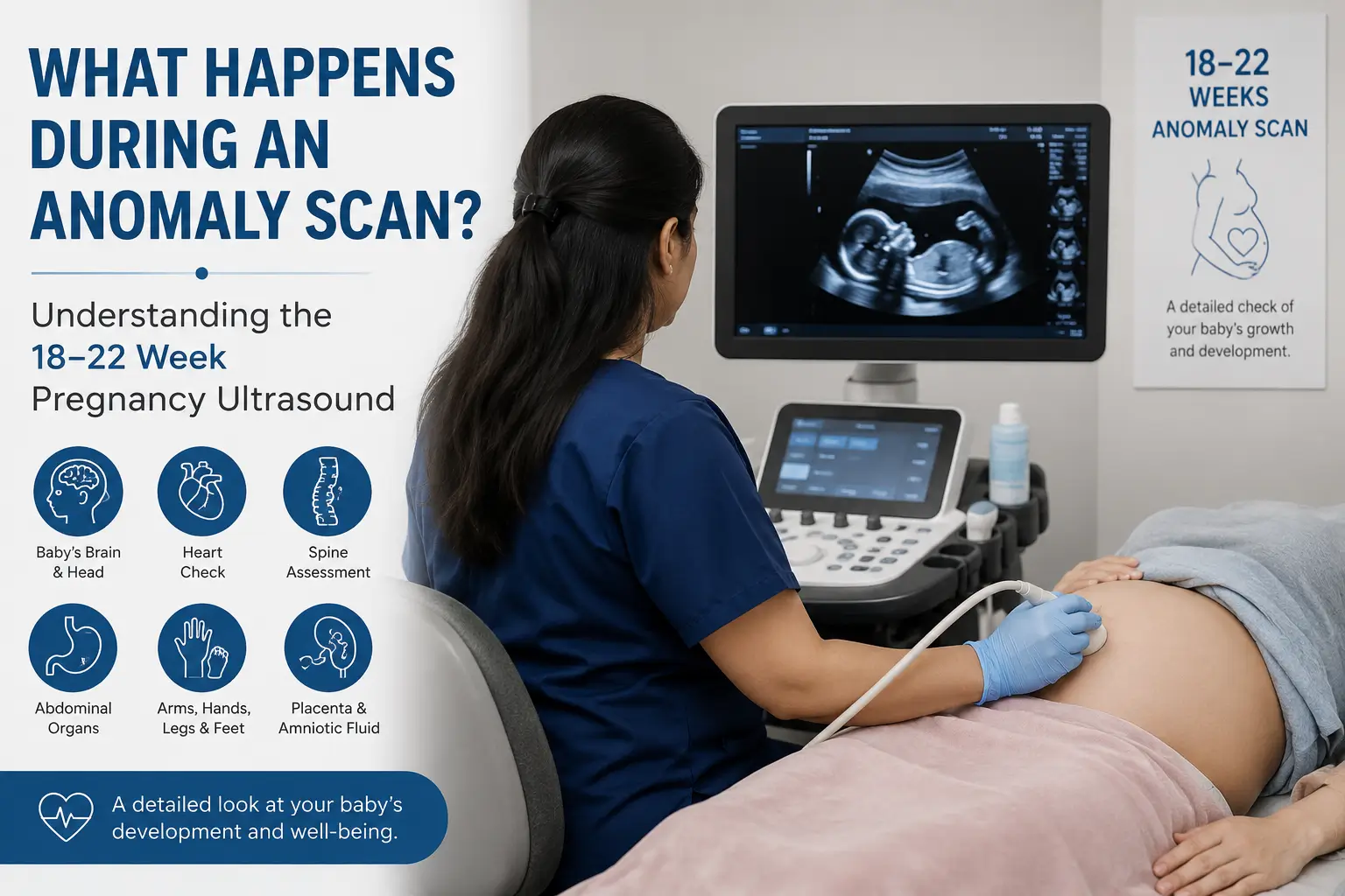
An anomaly scan is a detailed second-trimester ultrasound examination that is usually performed between 18 and 22 weeks of pregnancy. The purpose of the scan is to examine fetal anatomy as thoroughly as possible for that stage of development. During the examination, the sonographer or radiologist evaluates numerous structures throughout the baby's body. The scan helps assess development of the head, brain, face, spine, heart, abdomen, limbs and several internal organs. Although many parents associate the scan with obtaining clearer pictures of their baby, the primary purpose is medical. The examination provides information that may assist healthcare professionals in understanding how the pregnancy is progressing and whether fetal development appears consistent with expectations for that gestational age.
The timing of the anomaly scan is not arbitrary. By approximately 18 to 22 weeks of pregnancy, many fetal structures have developed sufficiently to allow detailed ultrasound assessment. At this stage, the baby is large enough for healthcare professionals to evaluate anatomical features while still small enough for most structures to be visualized during a single examination. Performing the scan too early may limit the ability to evaluate certain structures in adequate detail. Performing it much later may make comprehensive assessment more challenging because of fetal position, increasing size and other technical factors. For these reasons, the 18–22 week period is generally considered the optimal window for a detailed anatomy assessment.
One of the most common questions expectant parents ask is what exactly the sonographer is looking at during the examination. The answer is that the scan involves a systematic assessment of multiple areas of the baby's body. Rather than focusing on a single organ or measurement, the examination evaluates numerous anatomical structures from head to toe. The assessment may include evaluation of:
Each part of the examination contributes to a broader understanding of fetal development during the second trimester.
One of the most detailed parts of the anomaly scan involves assessment of the baby's head and developing brain. During the examination, healthcare professionals obtain specific ultrasound views that allow evaluation of important structures within the fetal head. Measurements of the head are also performed because these values help monitor growth and development. The examination is not limited to measuring size alone. The sonographer carefully examines anatomical structures that should be visible at this stage of pregnancy. This process requires patience, precision and appropriate fetal positioning. Parents may notice the examiner spending a considerable amount of time obtaining images of the head. This is completely normal and reflects the importance of collecting accurate information.
For many parents, seeing the baby's face becomes one of the most memorable moments of the anomaly scan. During the examination, healthcare professionals assess various facial structures including the profile and other anatomical features that can be visualized at this stage of development. Obtaining these images is not always straightforward. Babies frequently change position, move their hands near their face or turn away from the ultrasound probe. In some cases, the sonographer may need additional time to obtain suitable views. While parents often focus on the excitement of seeing facial features, the clinical purpose of these images is to assess development and anatomy as part of the overall examination.
The fetal spine is another important area evaluated during the anomaly scan. Healthcare professionals examine the spine from multiple angles and along different sections of the baby's back. Because the spine extends throughout much of the body, obtaining complete views may require adjustments in probe position and patience if the baby is moving. The assessment helps healthcare providers evaluate spinal development and visualize surrounding structures that are relevant during the examination. Parents often notice the sonographer moving the probe slowly across the abdomen while carefully observing the screen. Much of this time may be dedicated to obtaining clear spinal images.
Among all parts of the anomaly scan, assessment of the fetal heart is often one of the most technically demanding. The baby's heart is small, constantly moving and beating rapidly. Despite these challenges, ultrasound technology allows healthcare professionals to evaluate various aspects of cardiac anatomy and function. During the scan, the examiner obtains specific views of the heart and surrounding vessels. Blood flow may also be assessed in selected situations using Doppler techniques when clinically appropriate. Because cardiac assessment requires precision, parents should not be concerned if the sonographer spends a significant amount of time focusing on this area. Detailed evaluation is a routine and important part of the examination.
The anomaly scan also includes evaluation of several organs within the baby's abdomen. Structures that may be visualized include the stomach, kidneys, urinary bladder and other abdominal organs that can be assessed at this stage of pregnancy. The goal is not simply to confirm that these organs are present. Healthcare professionals also evaluate appearance, position and development according to established ultrasound assessment protocols. Many parents are surprised by how much information can be obtained from a routine ultrasound examination. Modern imaging technology allows remarkably detailed visualization of developing anatomy during pregnancy.
The anomaly scan includes careful evaluation of the baby's limbs. Healthcare professionals assess the arms, forearms, hands, thighs, lower legs and feet while obtaining measurements that contribute to growth assessment. This portion of the examination often provides some of the most recognizable images for parents. Tiny fingers, feet and limb movements may become visible during the scan, creating memorable moments for families. From a clinical perspective, however, these images also contribute important information regarding fetal anatomy and development. Because babies frequently move during the examination, obtaining suitable views may require patience and repeated attempts.
The anomaly scan focuses not only on the baby but also on structures that support the pregnancy. One of these structures is the placenta. During the examination, healthcare professionals assess placental position and appearance. Understanding placental location is important because it may influence pregnancy management and future monitoring. In many pregnancies, the placenta is positioned in a location that does not create concern. In others, follow-up assessment may occasionally be recommended. The placenta plays a vital role throughout pregnancy, supplying oxygen and nutrients that support fetal growth and development.
Amniotic fluid surrounds the baby throughout pregnancy and serves several important functions. During the anomaly scan, healthcare professionals assess amniotic fluid as part of the overall evaluation. Appropriate fluid volume contributes to fetal movement, development and protection within the uterus. Assessment of amniotic fluid is routinely incorporated into many pregnancy ultrasound examinations. The findings are interpreted together with other observations obtained during the scan. For this reason, the anomaly scan is not simply an examination of fetal anatomy. It is a broader assessment of multiple factors that contribute to pregnancy wellbeing.
Several measurements are obtained during the anomaly scan to help assess fetal growth. These measurements may include assessment of the head, abdomen and long bones. Together, they help healthcare professionals understand whether growth appears consistent with the expected gestational age. Growth measurements obtained during the anomaly scan also provide useful reference points for future examinations if additional pregnancy ultrasounds become necessary later. It is important to remember that ultrasound measurements are estimates rather than exact calculations. Healthcare professionals interpret these findings within the broader clinical context of the pregnancy.
Many parents associate the anomaly scan with learning the baby's gender. Because the examination occurs during the second trimester, visualization may be possible in some circumstances depending on fetal position and imaging conditions. However, the primary purpose of the anomaly scan is medical assessment rather than gender determination. In India, disclosure of fetal sex is prohibited under applicable laws governing prenatal diagnostic procedures. Healthcare providers follow these regulations strictly and do not disclose fetal gender during pregnancy ultrasound examinations. Parents should therefore approach the appointment with the understanding that the examination is intended to assess development, anatomy and pregnancy wellbeing.
One of the most common questions expectant parents ask before the appointment is how long the examination will take. The answer varies because every pregnancy is different. In many situations, the scan may take between twenty and forty-five minutes. Some examinations are completed more quickly, while others require additional time. Several factors influence the duration of the scan. These include fetal position, maternal body habitus, image quality and whether additional views are required. Parents should avoid comparing their appointment length with someone else's experience. A longer examination does not necessarily indicate a problem. In many cases, it simply means the sonographer is taking additional time to obtain complete images.
Many parents expect the anomaly scan to involve simply placing an ultrasound probe on the abdomen and taking a few pictures. In reality, the examination is a highly structured assessment that requires concentration, technical skill and careful image acquisition. Throughout the appointment, the sonographer or radiologist continuously adjusts the probe position to obtain specific views of different parts of the baby's body. Measurements are taken, images are recorded and various anatomical structures are assessed according to established ultrasound protocols. Parents often notice periods when the examiner becomes very focused on the screen. This is entirely normal. During these moments, the healthcare professional may be obtaining measurements, assessing anatomy or documenting important images for the medical record. The process is systematic and detailed because each image contributes to the overall assessment of fetal development.
For many families, the anomaly scan becomes one of the most memorable moments of pregnancy. Parents often experience a mixture of excitement, curiosity and nervousness before the examination begins. Once images appear on the screen, many are surprised by how clearly parts of the baby's body can be seen. Depending on fetal position, parents may observe movements such as stretching, turning, kicking or bringing hands toward the face. Some babies appear particularly active during the examination, while others seem content to remain asleep for much of the appointment. Although the experience can be emotional, it is important to remember that the scan remains a medical examination. The healthcare professional's primary focus is obtaining the information necessary to assess fetal development and pregnancy progress.
One concern occasionally raised by expectant parents is whether a longer examination indicates that something is wrong. In most situations, this assumption is incorrect. An anomaly scan may take additional time for many routine reasons. The baby may be facing away from the ultrasound probe, moving frequently or positioned in a way that makes certain structures difficult to visualize. Sometimes the sonographer simply needs additional images to complete the assessment properly. Obtaining high-quality diagnostic images often requires patience. Healthcare professionals prefer to spend extra time collecting accurate information rather than rushing through the examination. For this reason, a longer appointment should not automatically be viewed as a cause for concern.
Fetal position can significantly influence image quality during an anomaly scan. Occasionally, the baby may be lying in a position that prevents clear visualization of certain anatomical structures. When this occurs, the sonographer may ask the mother to change position, walk briefly or wait for the baby to move naturally. In many cases, these simple adjustments are enough to obtain the required views. Sometimes, however, additional imaging may be recommended if complete assessment cannot be achieved during the initial appointment. This does not necessarily indicate a problem. It simply reflects the practical challenges associated with imaging an active baby inside the uterus. Patience often becomes an important part of the process for both healthcare professionals and parents.
One of the most important facts parents should understand is that no ultrasound examination can identify every possible condition. Modern ultrasound technology provides remarkable diagnostic capability, but every medical test has limitations. Some conditions may be difficult to visualize, some may develop later in pregnancy and others may not be detectable using ultrasound techniques alone. The anomaly scan is designed to provide a detailed assessment of fetal anatomy at a specific stage of development. While it can provide valuable information, it cannot guarantee identification of every possible concern. Healthcare professionals interpret findings within the limitations of the technology, fetal position and individual pregnancy circumstances. Understanding these limitations helps establish realistic expectations regarding the purpose and capabilities of the examination.
Most anomaly scans are completed without identifying significant concerns. However, there are occasions when an observation requires additional evaluation. If further assessment is considered necessary, healthcare providers will explain the findings and discuss appropriate next steps. Depending on the situation, recommendations may include follow-up ultrasound examinations, specialist consultation or additional monitoring during pregnancy. Receiving unexpected information can understandably create anxiety for parents. It is important to remember that many findings require further evaluation before conclusions can be reached. The purpose of additional assessment is to gather more information and support informed clinical decision-making rather than to assume the presence of a serious problem.
Preparation requirements for an anomaly scan may vary depending on local protocols and the stage of pregnancy. Patients should always follow the instructions provided by their healthcare provider or diagnostic centre. Some facilities may recommend arriving with a partially filled bladder, while others may not require any special preparation. Wearing comfortable clothing can make the examination more convenient. It is also helpful to arrive a little before the scheduled appointment time to complete any necessary paperwork. When preparation instructions are unclear, contacting the diagnostic centre in advance is always advisable.
Bringing relevant documents can help ensure a smooth and efficient appointment. Healthcare professionals may benefit from reviewing previous pregnancy ultrasounds, antenatal records and referral notes when available. Useful items may include:
Maintaining organized pregnancy records can be valuable throughout prenatal care and may assist healthcare providers in understanding the progression of the pregnancy over time.
No. The examination is generally painless. The ultrasound probe is moved across the abdomen while images are obtained using sound-wave technology.
During this period, many fetal structures have developed sufficiently to allow detailed anatomical assessment while still being suitable for comprehensive ultrasound evaluation.
No. While the examination provides extensive information, every medical test has limitations and not all conditions can be identified through ultrasound.
The duration varies, but many anomaly scans take approximately twenty to forty-five minutes depending on fetal position and imaging requirements.
Healthcare providers may recommend follow-up imaging or specialist consultation if further evaluation is considered appropriate.
The anomaly scan represents one of the most detailed and informative ultrasound examinations performed during pregnancy. Conducted between 18 and 22 weeks, it provides healthcare professionals with an opportunity to carefully assess fetal anatomy, monitor development and gather valuable information about the progress of the pregnancy. For parents, the appointment often becomes a memorable milestone. It offers a chance to see detailed images of the baby while gaining a deeper understanding of how pregnancy is progressing. At the same time, it serves an important clinical purpose by helping healthcare professionals evaluate numerous aspects of fetal development. Although no ultrasound examination can provide every answer, the anomaly scan remains a valuable component of modern prenatal care. Understanding what happens during the examination can help expectant parents approach the appointment with greater confidence, realistic expectations and peace of mind.
Medical content published by Orbit Diagnostics & Healthcare is reviewed periodically to help ensure accuracy, relevance and alignment with accepted diagnostic imaging practices.
Lead Medical Reviewer
Dr. Nidha Nazir
MBBS, MD Radiology
Consultant Radiologist
Note: Content is intended for educational purposes and does not replace professional medical advice, diagnosis or treatment.