Heart Evaluation & Echocardiography

Echo Test in Srinagar

Disclaimer: This page is intended to support patient understanding and should not replace professional medical advice, diagnosis or treatment.

Echocardiography For Heart Function, Heart Valves & Cardiac Assessment

If your doctor has advised an Echo test, you probably have questions. Many patients want to know what the test involves, whether it is painful, how long it takes, whether any preparation is required and when the report will be available.

At Orbit Diagnostics & Healthcare, Srinagar, Echo (Echocardiography) services are available to help evaluate heart structure, heart function, heart valves and blood flow through the heart. The examination is non-invasive, does not use radiation and is commonly requested by cardiologists, physicians and other healthcare professionals when assessing heart-related symptoms and conditions.

Echo Test At A Glance

  • Test Name: Echocardiography (Echo)
  • Purpose: Evaluation of heart structure and function
  • Radiation: No radiation exposure
  • Pain: Usually painless
  • Typical Duration: Approximately 15–30 minutes
  • Preparation: Usually minimal
  • Report: According to cardiology reporting workflow
  • Location: Baghat Chowk, Srinagar

Echo Test Cost Comparison Overview

The cost of an Echo test can vary depending on the city, healthcare facility, type of Echocardiography being performed and the complexity of the examination. The table below provides a general overview of commonly reported price ranges across India.

Test Type Average Cost Range (INR) Premium Hospital / Metro City Range (INR)
Adult Echocardiogram (Standard 2D Echo) ₹1,200 – ₹3,500 ₹4,000 – ₹5,000
Fetal Echocardiogram ₹2,000 – ₹5,000 ₹6,000 – ₹10,000+

Estimated Echo Test Costs In Major Indian Cities

The following figures are approximate examples of commonly reported market ranges and may vary between healthcare providers.

City Estimated Adult Echo Cost Estimated Fetal Echo Cost
Delhi / NCR ₹1,500 – ₹3,000 ₹3,000 – ₹6,000
Mumbai / Thane ₹1,800 – ₹3,500 ₹3,500 – ₹7,000
Bengaluru ₹1,500 – ₹3,200 ₹2,500 – ₹5,500
Hyderabad ₹1,200 – ₹2,500 ₹1,500 – ₹5,000
Kolkata ₹1,500 – ₹2,800 ₹2,500 – ₹5,000

Actual charges can vary depending on healthcare provider, equipment used, reporting requirements, specialist involvement and regional market conditions. Patients are encouraged to contact the clinic directly for the latest pricing information and appointment availability.

Why Do Echo Test Prices Vary?

City & Location

Charges in major metropolitan cities such as Delhi, Mumbai, Bengaluru and Chennai are often higher than those in smaller cities and regional healthcare centres. Local operating costs, demand and healthcare infrastructure can influence pricing.

Type Of Healthcare Facility

Independent diagnostic centres and specialised imaging facilities may have different pricing structures compared with large corporate hospital networks. Hospital-based services often include additional operational and administrative costs.

Complexity Of The Examination

A routine adult 2D Echo is generally less expensive than advanced cardiac imaging procedures. Examination complexity, reporting requirements and specialist involvement can affect the final charge.

Advanced Echocardiography Procedures

Adult Cardiac Imaging

  • Standard 2D Echo With Color Doppler: Routine evaluation of heart chambers, valves and blood flow.
  • Stress Echocardiography: Performed before and after exercise or pharmacological stress to evaluate heart function under increased workload.
  • Transesophageal Echocardiography (TEE): A specialised examination performed using an ultrasound probe inserted through the oesophagus, often providing highly detailed cardiac images.

Advanced examinations such as Stress Echo and TEE may cost significantly more than a routine Echocardiogram and can range from approximately ₹6,000 to ₹12,000 or more depending on the healthcare facility and clinical requirements.

Fetal Echocardiography

  • Standard fetal heart assessment.
  • Detailed fetal cardiac evaluation.
  • Twin pregnancy cardiac assessment.
  • Advanced 3D/4D fetal cardiac imaging where available.

The complexity of the fetal examination, gestational age and reporting requirements may influence the overall cost.

What Happens During An Echo Test?

One of the most common concerns patients have is uncertainty about what will happen during the examination. Understanding the process often makes the experience much more comfortable.

01

Arrival & Registration

Patients arrive at the clinic, complete registration procedures and provide any referral documents or previous reports.

02

Preparation

The healthcare team explains the procedure and helps prepare the patient for the examination.

03

Ultrasound Examination

A specialised cardiac ultrasound probe is placed on different areas of the chest to obtain images of the heart.

04

Image Recording

Images and measurements are recorded for clinical review and interpretation.

05

Cardiology Review

The findings are reviewed according to reporting requirements and clinical workflow.

06

Report Preparation

The report is prepared and made available according to clinic procedures.

Does An Echo Test Hurt?

For most patients, Echocardiography is painless. The ultrasound probe is moved gently across the chest while images are recorded. Some patients may feel mild pressure at certain positions, but the examination generally does not cause pain.

Is Echo Safe?

Echo uses ultrasound technology rather than radiation. It is widely used for heart assessment and is considered a routine diagnostic investigation when medically indicated.

How Long Does An Echo Test Take?

The duration varies depending on the reason for the examination and the information required. Many Echo examinations are completed within approximately 15 to 30 minutes, although individual cases may vary.

How To Prepare For An Echo Test

One advantage of Echocardiography is that it usually requires very little preparation. Most patients can continue their normal daily activities before the examination. However, bringing the right information and arriving prepared can help make the visit smoother and more productive.

Before You Arrive

  • Carry your doctor's prescription or referral, if available.
  • Bring previous Echo reports, ECG reports or cardiac investigations.
  • Carry a list of medications you are currently taking.
  • Reach the clinic approximately 10–15 minutes before your appointment.
  • Wear comfortable clothing that allows easy access to the chest area.
  • Inform the healthcare team about any recent heart procedures or hospital admissions.
  • Bring any relevant medical records that may help with interpretation.

Common Preparation Questions

Is Fasting Required?

For a routine transthoracic Echo, fasting is generally not required. Patients can usually eat and drink normally unless specifically advised otherwise by their doctor.

Can I Drink Water Before The Test?

In most cases, yes. Drinking water is usually permitted before a routine Echo examination.

Can I Take My Regular Medicines?

Patients should generally continue medications exactly as advised by their treating doctor unless instructed differently by a healthcare professional.

Do I Need To Stop Any Medication?

Do not stop prescribed medicines unless your doctor specifically instructs you to do so. If you are uncertain, discuss your medication list with your healthcare provider before the appointment.

What Should I Bring To My Appointment?

Item Why It Helps
Doctor's Prescription Provides clinical background and reason for examination.
Previous Echo Reports Allows comparison with earlier findings.
ECG Reports Provides additional cardiac information.
Medication List Helps healthcare professionals understand current treatment.
Relevant Medical Records May assist with interpretation and continuity of care.

Do I Need A Doctor's Prescription For An Echo Test?

Many patients arrive with a referral from a cardiologist, physician or another treating doctor. The referral helps provide important clinical information regarding symptoms, medical history and the reason the examination has been requested.

If you already have a prescription, it is helpful to bring it with you. The referral often allows the reporting doctor to better understand the clinical question that needs to be addressed.

Patients who are unsure whether a prescription is required should contact the clinic before their visit. The team can explain current appointment requirements and guide patients regarding documentation that may be needed.

When Will My Echo Report Be Available?

Routine Reporting

Many Echo examinations can be reported on the same day according to workflow and reporting requirements.

Review Process

Images and measurements may require review before final report preparation. Reporting timelines can vary depending on clinical circumstances.

Urgent Cases

Where clinically necessary, treating doctors may receive important findings according to established communication procedures.

Patients are encouraged to ask about expected reporting timelines at the time of appointment booking or registration.

Why Patients Choose Orbit Diagnostics For Echo Tests

Dedicated Cardiac Assessment

Echo examinations focus on evaluating heart structure, heart valves, blood flow and cardiac function.

Modern Ultrasound Technology

Cardiac imaging is supported by contemporary ultrasound equipment designed for diagnostic imaging applications.

Qualified Medical Oversight

Examinations follow established clinical workflows and reporting processes.

Convenient Srinagar Location

Located at Baghat Chowk with convenient access from multiple areas of Srinagar and surrounding districts.

Appointment Assistance

Patients can contact the clinic regarding appointments, preparation requirements and availability.

Integrated Diagnostic Services

Patients may access multiple diagnostic services under one roof including ECG, Holter Monitoring, ABPM and laboratory investigations.

Why Has My Doctor Recommended An Echo Test?

Many patients arrive with a referral for Echocardiography but are unsure why the examination has been advised. The reason varies from one patient to another. The test helps doctors obtain important information about heart structure and function that cannot be obtained through symptoms alone.

Chest Pain

An Echo may be recommended when evaluating heart-related causes of chest discomfort.

Shortness Of Breath

Breathlessness can sometimes be related to heart function and may require further evaluation.

High Blood Pressure

Long-standing hypertension may affect the heart and doctors may request Echocardiography as part of assessment.

Heart Murmur

Heart valve function can often be assessed through Echocardiography.

Palpitations

Patients experiencing irregular or rapid heartbeats may be referred for Echo evaluation.

Known Heart Disease

Echocardiography is frequently used to monitor previously diagnosed cardiac conditions.

A Common Concern We Hear From Patients

Many people become anxious when a doctor recommends an Echo test because they assume it means a serious heart problem has already been identified. In reality, Echocardiography is frequently requested simply because a doctor wants additional information about how the heart is functioning.

The examination is often used to investigate symptoms, monitor existing conditions, evaluate heart murmurs, assess blood pressure-related changes or provide reassurance when symptoms need further assessment. An Echo request does not automatically mean a major heart disease has been found.

If your doctor has advised an Echo test, the best approach is usually to complete the examination and review the findings with the healthcare professional managing your care.

What Is An Echo Test?

An Echo test, also known as Echocardiography, is an ultrasound examination of the heart. Just as ultrasound can create images of organs inside the abdomen or during pregnancy, Echocardiography uses sound waves to create real-time images of the heart while it is beating.

The examination allows doctors to observe how the heart chambers move, how the valves open and close, how effectively blood flows through the heart and whether there are structural or functional abnormalities that may require further medical attention.

Unlike X-rays or CT scans, an Echo test does not use ionising radiation. A small ultrasound probe is placed on the chest and images are generated using sound-wave technology. Because the examination is non-invasive and generally comfortable, it is one of the most commonly performed cardiac investigations worldwide.

Doctors may request an Echo test when evaluating symptoms such as chest discomfort, breathlessness, palpitations, swelling of the legs, heart murmurs, dizziness, uncontrolled blood pressure or suspected heart disease. It may also be recommended as part of follow-up care for individuals with previously diagnosed cardiac conditions.

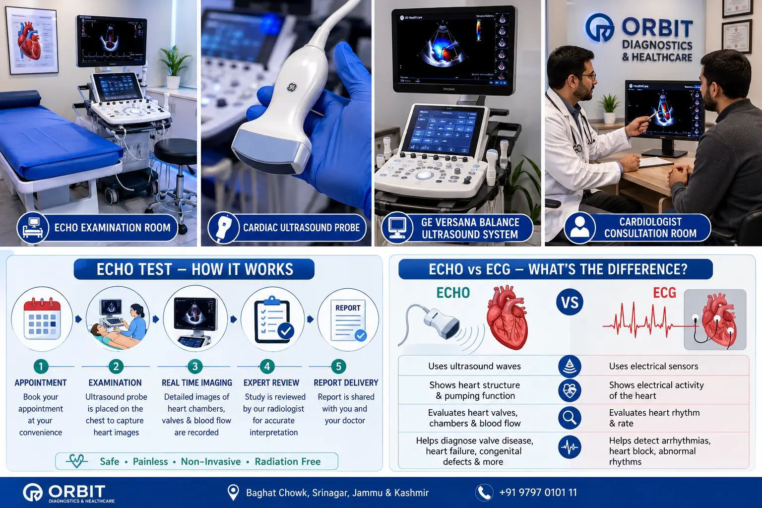
Echo Test in Srinagar infographic showing Echo examination room, cardiac ultrasound probe, GE Versana Balance ultrasound machine, cardiologist consultation room, Echo test workflow and Echo versus ECG comparison
Echo Test (Echocardiography) Patient Information Guide: This educational infographic explains the complete Echo test journey, including the examination environment, cardiac ultrasound technology, GE Versana Balance imaging system, cardiologist consultation process, step-by-step Echo workflow and the key differences between Echocardiography and ECG. It helps patients understand what to expect before, during and after an Echo test at Orbit Diagnostics & Healthcare, Srinagar.

What Can An Echo Test Help Evaluate?

An Echo test provides valuable information about how the heart looks and how it functions. While the final interpretation depends on clinical assessment and medical review, Echocardiography can help doctors evaluate a wide range of heart-related conditions.

Condition Or Concern How Echo May Help
Heart Valve Disease Helps evaluate how heart valves open, close and allow blood to flow.
Heart Failure Assesses heart pumping function and chamber performance.
Cardiomyopathy May help identify changes in heart muscle structure and function.
Congenital Heart Conditions Can assist in evaluating certain structural abnormalities present from birth.
Pulmonary Hypertension May provide information regarding heart pressures and blood flow patterns.
Pericardial Effusion Can detect fluid surrounding the heart.
Heart Chamber Enlargement Assesses chamber size and structural changes.
Reduced Heart Function Helps determine how effectively the heart is pumping blood.

Understanding A Typical Echo Report

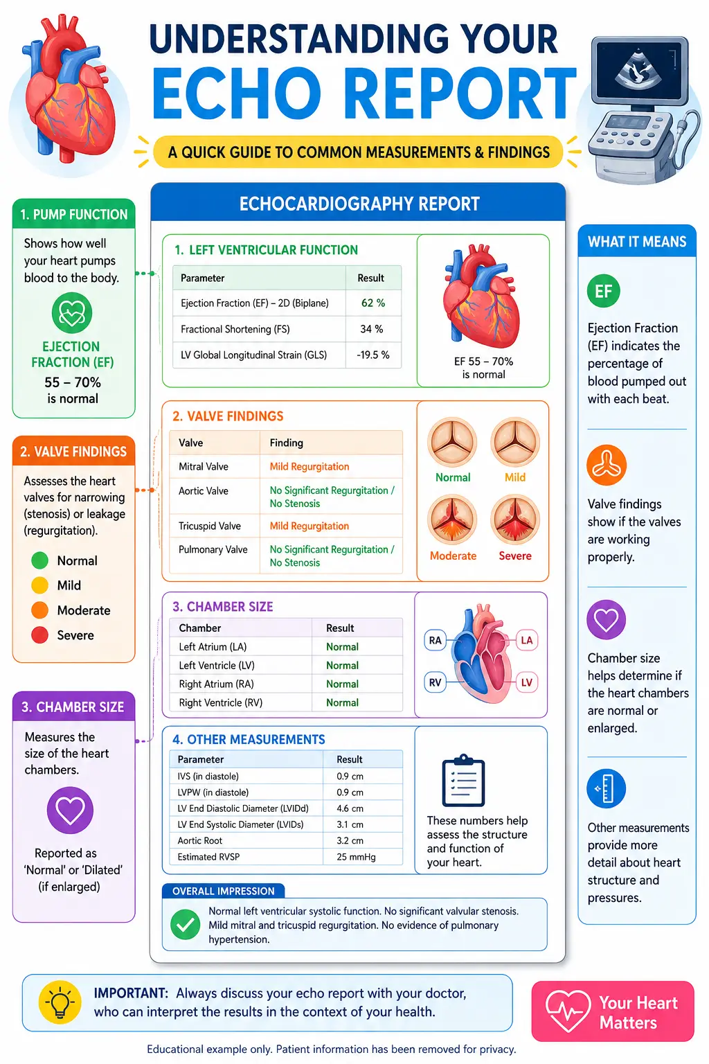
Many patients receive an Echo report but are unsure what the medical terms mean. The example below shows common measurements and findings that may appear on a routine Echocardiography report. This sample is provided for educational purposes only and does not represent an actual patient.

Parameter Sample Finding What It Means
LVEF (Left Ventricular Ejection Fraction) 60% Measures how effectively the heart pumps blood.
LV End Diastolic Diameter (LVEDD) 48 mm Measurement of left ventricular size.
LV End Systolic Diameter (LVESD) 31 mm Measurement of ventricular size after contraction.
Left Atrium Normal Size Assessment of left atrial dimensions.
Right Ventricle Normal Function Evaluation of right-sided heart performance.
Mitral Valve No Significant Abnormality Assessment of valve structure and function.
Aortic Valve Normal Opening Evaluation of blood flow from the heart.
Tricuspid Valve Trace Physiological Regurgitation A commonly reported minor finding.
Pericardium No Effusion No excess fluid surrounding the heart.
Pulmonary Artery Pressure Normal Assessment of pressure within pulmonary circulation.

Sample Impression

Normal left ventricular size and systolic function with an estimated ejection fraction of 60%. No significant valvular abnormality identified. No pericardial effusion. Overall findings consistent with a structurally normal heart.

This sample report is provided solely for educational purposes. Actual Echo findings vary from patient to patient and must always be interpreted by the reporting doctor in the context of symptoms, examination findings and medical history.

Sample Echocardiography report showing ejection fraction, valve findings and cardiac measurements
Educational example of an Echo report showing common cardiac measurements and findings. Patient information has been removed for privacy.

What Is The Difference Between Echo And ECG?

Many patients assume that ECG and Echo are the same test. In reality, they provide different types of information and are often used together when assessing heart health.

Feature Echo Test ECG
Evaluates Heart Structure ✓ Yes ✗ No
Evaluates Heart Function ✓ Yes Limited
Shows Electrical Activity ✗ No ✓ Yes
Uses Ultrasound ✓ Yes ✗ No
Provides Real-Time Images ✓ Yes ✗ No
Commonly Used Together ✓ Yes ✓ Yes

In many situations, doctors request both ECG and Echo because each test provides different information about heart health.

Cardiologist Oversight & Clinical Review

An Echo test is more than simply obtaining images of the heart. The value of the examination comes from proper acquisition of images, accurate measurements and clinical interpretation of the findings.

Echo examinations are performed according to established cardiac imaging workflows. Findings are reviewed within the appropriate reporting process so that the information can be used by the treating doctor when making clinical decisions.

Patients often focus on the machine performing the examination, but equally important is the professional interpretation of the images and how those findings are integrated into the patient's overall medical assessment.

Echo Technology & Cardiac Imaging Equipment

The quality of an Echo examination depends on more than simply obtaining images. Accurate cardiac assessment requires suitable imaging technology, proper examination technique and professional interpretation of the findings.

At Orbit Diagnostics & Healthcare, cardiac imaging examinations are performed using modern ultrasound technology capable of producing detailed real-time images of the heart and blood flow patterns.

During an Echo test, the cardiac ultrasound probe captures images from different angles around the chest, allowing assessment of heart chambers, valves, pumping function and blood flow dynamics.

Clear imaging helps support accurate measurements and clinical interpretation, which ultimately assists treating doctors in making informed healthcare decisions.

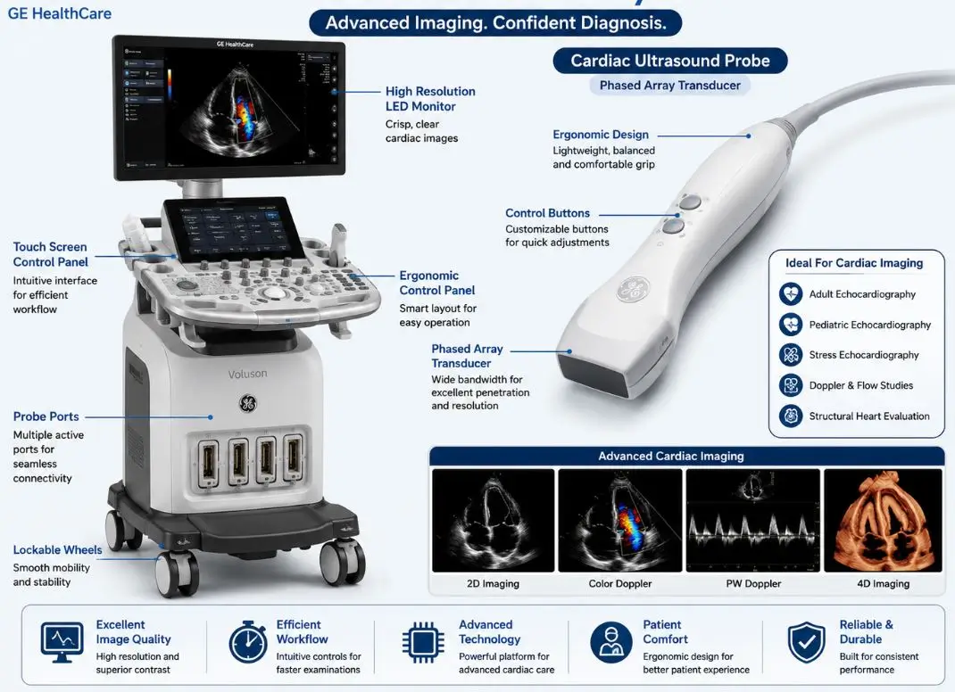
Cardiac ultrasound probe used for Echocardiography at Orbit Diagnostics & Healthcare Srinagar
Cardiac ultrasound probe used for Echocardiography and heart function assessment.

GE Versana Balance Ultrasound Platform

Orbit Diagnostics & Healthcare uses the GE Versana Balance ultrasound platform, a modern imaging system designed to support a wide range of diagnostic ultrasound applications including cardiac imaging, abdominal imaging, pregnancy ultrasound and Doppler studies.

For Echo examinations, specialised cardiac imaging capabilities assist with evaluation of heart anatomy, movement and blood flow patterns. Modern ultrasound technology helps provide the image quality required for detailed cardiac assessment.

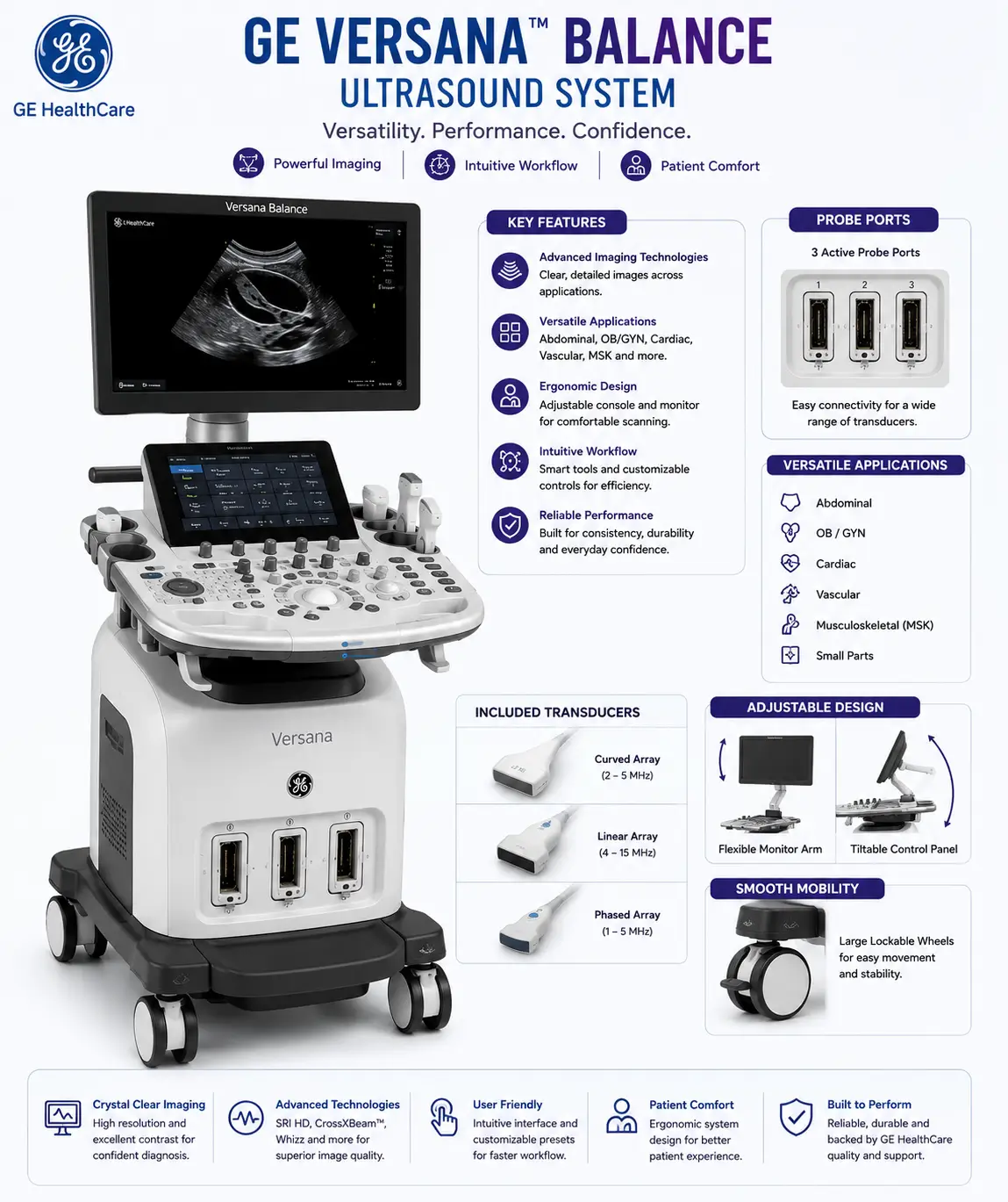
GE Versana Balance Ultrasound Technology at Orbit Diagnostics & Healthcare Srinagar
GE Versana Balance Ultrasound System: Advanced ultrasound technology supporting Echocardiography, Doppler studies, pregnancy imaging, abdominal ultrasound and general diagnostic examinations.

Why Patients Travel From Across Kashmir For Echo Tests

While many patients visiting Orbit Diagnostics & Healthcare are from Srinagar, a significant number travel from surrounding districts when referred for cardiac investigations and diagnostic services.

Some patients are sent by their family doctors for heart evaluation. Others are referred by physicians, cardiologists or healthcare facilities seeking Echocardiography as part of ongoing medical assessment.

The clinic is accessible for patients travelling from Budgam, Ganderbal, Pulwama, Shopian, Kulgam, Anantnag, Baramulla, Bandipora, Kupwara and other areas across the Kashmir Valley.

For patients travelling longer distances, appointment scheduling can help reduce waiting times and provide clarity regarding availability before arrival.

Areas Commonly Served

Patients regularly visit Orbit Diagnostics & Healthcare from various parts of Srinagar and neighbouring districts.

  • Srinagar
  • Baghat
  • Barzulla
  • Rawalpora
  • Natipora
  • Chanapora
  • Hyderpora
  • Nowgam
  • Rangreth
  • Humhama
  • Budgam
  • Ganderbal
  • Pulwama
  • Shopian
  • Kulgam
  • Anantnag
  • Baramulla
  • Bandipora
  • Kupwara
  • Handwara
  • Gulmarg Region

Medical Quality, Professional Standards & Patient Confidence

A heart test can naturally create anxiety. Many patients arrive concerned about what the examination might reveal, while others simply want reassurance that their symptoms are being properly evaluated.

Our goal is to provide a professional diagnostic environment where patients understand what to expect before, during and after their examination. Clear communication, appropriate appointment guidance and structured reporting processes are all part of delivering a positive patient experience.

Whether the Echo examination has been requested for chest discomfort, shortness of breath, blood pressure monitoring, follow-up of an existing cardiac condition or general evaluation, every patient deserves accurate information and respectful care throughout the diagnostic process.

Frequently Asked Questions About Echo Tests

The questions below are based on common concerns raised by patients before scheduling an Echocardiography examination.

What is an Echo test?

An Echo test, or Echocardiography, is an ultrasound examination that creates real-time images of the heart and helps doctors evaluate heart structure, valves and pumping function.

Is an Echo test painful?

No. Most patients find the examination comfortable. The ultrasound probe is moved over the chest and typically causes little more than mild pressure.

How long does an Echo test take?

Many Echo examinations are completed within approximately 15 to 30 minutes, although duration may vary according to the clinical situation.

Is fasting required before an Echo test?

Routine Echocardiography generally does not require fasting unless specific instructions have been provided by your doctor.

Can I drink water before the examination?

Yes. Drinking water is generally permitted before a routine Echo examination.

Can I take my regular medicines?

Patients should normally continue prescribed medicines unless their doctor advises otherwise.

Do I need an appointment?

Appointments are recommended because they help minimise waiting times and allow the clinic to provide preparation guidance where required.

Can I come without a doctor's prescription?

Patients are encouraged to bring a referral or prescription whenever available. If you are unsure about requirements, please contact the clinic before your visit.

What should I bring for my Echo test?

Bring your prescription, previous Echo reports, ECG reports, medication list and any relevant cardiac records.

Does Echo use radiation?

No. Echocardiography uses ultrasound waves and does not expose patients to ionising radiation.

Can Echo detect heart valve problems?

Echo is commonly used to assess how heart valves open, close and regulate blood flow.

Can Echo evaluate heart pumping function?

Yes. One of the most important uses of Echocardiography is assessing how effectively the heart pumps blood.

Can Echo detect heart failure?

Echo may provide information that helps doctors evaluate heart failure and related cardiac conditions.

Can children undergo Echo examinations?

Depending on the clinical requirement, Echocardiography may also be performed for children when advised by the treating doctor.

Is Echo safe during pregnancy?

Because Echo uses ultrasound technology rather than radiation, it is generally considered a safe imaging technique when medically indicated.

When will my report be available?

Many examinations can be reported on the same day, although timing may vary according to workflow and clinical review requirements.

What is the difference between ECG and Echo?

ECG evaluates the electrical activity of the heart, while Echo provides images of the heart's structure and function. Doctors often use both investigations together.

Can Echo detect blocked arteries?

Echo can provide important information about heart function and blood flow, but evaluation of coronary artery blockages may require additional investigations depending on the clinical situation.

How often should an Echo be repeated?

The frequency depends on your medical condition and your doctor's recommendations.

Where can I get an Echo test in Srinagar?

Orbit Diagnostics & Healthcare provides Echo testing services at Baghat Chowk, Srinagar with appointment support and diagnostic imaging facilities.

Medical Review and Content Oversight

Medical content published by Orbit Diagnostics & Healthcare is reviewed periodically to help ensure accuracy, relevance and alignment with accepted diagnostic imaging practices.

Lead Medical Reviewer
Dr. Nidha Nazir
MBBS, MD Radiology
Consultant Radiologist

Note: Content is intended for educational purposes and does not replace professional medical advice, diagnosis or treatment.

Need An Echo Test In Srinagar?

Whether your doctor has advised an Echo examination, you are seeking follow-up evaluation for an existing heart condition or you have questions about appointment availability, our team is available to help.

Contact Orbit Diagnostics & Healthcare for appointment guidance, preparation information and scheduling support.