Obstetric & Pregnancy Ultrasound in Srinagar
Disclaimer: This page is intended to support patient understanding and should not replace professional medical advice, diagnosis or treatment.
Pregnancy is a journey marked by important developmental milestones for both mother and baby. Obstetric and pregnancy ultrasound examinations help monitor fetal growth, assess wellbeing, evaluate maternal structures, and provide valuable information throughout pregnancy. At Orbit Diagnostics & Healthcare, Srinagar, pregnancy ultrasound examinations are performed using modern ultrasound technology under the supervision of experienced radiology professionals.
Pregnancy Ultrasound At A Glance
Pregnancy ultrasound uses high-frequency sound waves to create real-time images of the developing baby and maternal structures. It plays an important role in confirming pregnancy, estimating gestational age, assessing fetal development, monitoring growth, evaluating the placenta, and assessing fetal wellbeing throughout pregnancy.
| Feature | Details |
|---|---|
| Service | Obstetric & Pregnancy Ultrasound |
| Patient Group | Pregnant Women |
| Imaging Method | Ultrasound Sound Waves |
| Radiation Exposure | None |
| Pregnancy Stages Covered | First, Second & Third Trimester |
| Common Examinations | Viability Scan, NT Scan, Anomaly Scan, Growth Scan, Doppler Assessment |
| Reporting | Consultant Radiologist |
| Location | Orbit Diagnostics & Healthcare, Srinagar |
Orbit Diagnostics is an established Ultrasound Clinic in Srinagar providing pregnancy, abdominal, pelvic, vascular and general ultrasound examinations.
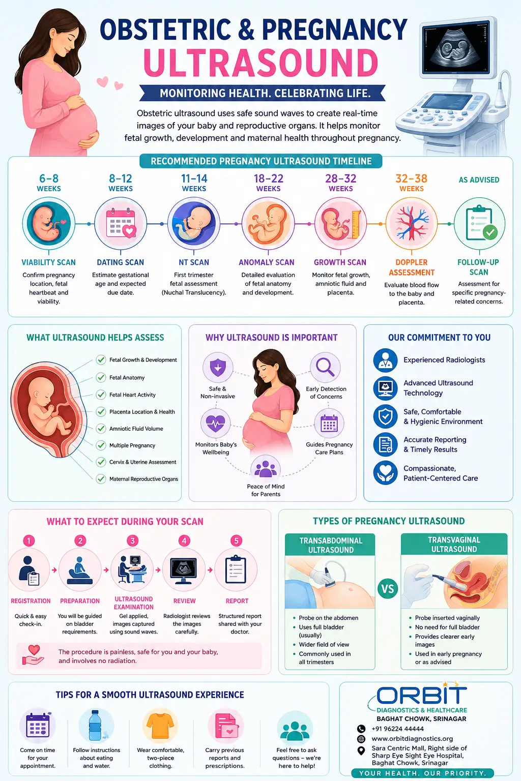
Recommended Pregnancy Ultrasound Timeline
Different ultrasound examinations may be recommended at different stages of pregnancy. The exact schedule may vary depending on individual clinical circumstances and recommendations from the treating obstetrician.
| Pregnancy Week | Common Scan | Main Purpose |
|---|---|---|
| 6–8 Weeks | Viability Scan | Confirm pregnancy location, fetal heartbeat and viability |
| 8–12 Weeks | Dating Scan | Estimate gestational age and expected due date |
| 11–14 Weeks | NT Scan | First trimester fetal assessment |
| 18–22 Weeks | Anomaly Scan | Detailed fetal anatomical evaluation |
| 28–32 Weeks | Growth Scan | Monitor fetal growth and development |
| 32–38 Weeks | Doppler Assessment | Evaluate blood flow when clinically indicated |
| As Advised | Follow-Up Scan | Assessment for specific pregnancy-related concerns |
Pregnancy Week Guide: Common Ultrasound Milestones
The timing of ultrasound examinations varies according to individual clinical circumstances and your doctor's recommendations. The table below provides a general overview of common pregnancy imaging milestones.
| Pregnancy Week | Common Assessment | Purpose |
|---|---|---|
| 6–8 Weeks | Viability Scan | Confirm pregnancy location and fetal heartbeat |
| 8–10 Weeks | Dating Assessment | Estimate gestational age |
| 11–14 Weeks | NT Scan | First trimester fetal assessment |
| 15–17 Weeks | Follow-Up Assessment (if advised) | Individual clinical requirements |
| 18–22 Weeks | Anomaly Scan | Detailed fetal anatomical evaluation |
| 23–27 Weeks | Follow-Up Assessment (if advised) | Monitoring specific clinical concerns |
| 28–32 Weeks | Growth Scan | Growth and development monitoring |
| 32–36 Weeks | Doppler Assessment (when indicated) | Blood flow evaluation |
| 36–40 Weeks | Late Pregnancy Assessment | Position, growth and wellbeing assessment |
Questions You May Wish To Discuss With Your Doctor Before A Pregnancy Ultrasound
Every pregnancy is unique. Discussing the purpose of the examination with your healthcare provider can help you better understand how the ultrasound contributes to your pregnancy care.
| Question | Why It May Be Helpful |
|---|---|
| Why has this ultrasound been recommended? | Understanding the purpose of the examination |
| What information will this scan provide? | Clarifies expectations regarding the assessment |
| Do I need any preparation before the scan? | Helps improve examination readiness |
| Will additional scans be required later? | Provides understanding of future monitoring |
| Are there any specific concerns being evaluated? | Improves awareness of clinical objectives |
| Should I bring previous reports? | Allows comparison with earlier findings |
| Will Doppler assessment be required? | Provides insight into additional imaging needs |
| When should I return for the next scan? | Helps plan ongoing pregnancy care |
Quick Pregnancy Scan Reference Guide
The following table summarizes the most commonly performed obstetric ultrasound examinations.
| Scan | Typical Timing | Main Focus |
|---|---|---|
| Viability Scan | 6–8 Weeks | Pregnancy confirmation and heartbeat |
| Dating Scan | 8–12 Weeks | Gestational age estimation |
| NT Scan | 11–14 Weeks | First trimester fetal assessment |
| Anomaly Scan | 18–22 Weeks | Detailed anatomy evaluation |
| Growth Scan | 28–32 Weeks | Growth monitoring |
| Doppler Study | As Recommended | Blood flow assessment |
| Late Pregnancy Scan | 36+ Weeks | Position and wellbeing assessment |
What Is Obstetric & Pregnancy Ultrasound?
Obstetric ultrasound is a medical imaging examination used during pregnancy to assess the developing fetus and maternal reproductive structures. Unlike X-rays or CT scans, ultrasound uses sound waves rather than ionizing radiation, making it a commonly used imaging method during pregnancy.
Patients seeking routine Pregnancy Ultrasound in Srinagar may undergo different examinations depending on the stage of pregnancy and their doctor's recommendations.
Pregnancy ultrasound examinations may be performed during the first, second, and third trimesters depending on the clinical requirements of the pregnancy. These examinations provide important information regarding fetal growth, fetal position, placental location, amniotic fluid volume, and overall pregnancy progression.
Modern ultrasound systems can provide detailed visualization of fetal anatomy and may assist healthcare providers in monitoring pregnancy development and planning ongoing care.
Why Pregnancy Ultrasounds Are Important
Pregnancy ultrasound examinations provide valuable information throughout pregnancy and help clinicians monitor both maternal and fetal wellbeing.
| Area Assessed | Clinical Information Obtained |
|---|---|
| Pregnancy Confirmation | Confirmation of intrauterine pregnancy and viability |
| Fetal Heart Activity | Assessment of fetal cardiac activity |
| Gestational Age | Estimation of pregnancy age and expected delivery date |
| Fetal Growth | Monitoring growth parameters over time |
| Fetal Anatomy | Assessment of developing organs and structures |
| Placenta | Evaluation of placental position and appearance |
| Amniotic Fluid | Assessment of fluid volume surrounding the fetus |
| Fetal Position | Monitoring fetal presentation and position |
| Multiple Pregnancy | Evaluation of twin or multiple gestations |
| Blood Flow | Doppler assessment when clinically indicated |
Pregnancy Milestones And Corresponding Ultrasound Assessments
Different ultrasound examinations help assess specific milestones throughout pregnancy.
| Pregnancy Milestone | Common Ultrasound Assessment |
|---|---|
| Confirmation of Pregnancy | Viability Scan |
| Detection of Fetal Heartbeat | Early Pregnancy Ultrasound |
| Due Date Estimation | Dating Scan |
| Early Fetal Development Assessment | NT Scan |
| Detailed Anatomy Evaluation | Anomaly Scan |
| Monitoring Growth | Growth Scan |
| Placental Function Assessment | Doppler Evaluation |
| Assessment Before Delivery | Late Pregnancy Ultrasound |
Pregnancy Ultrasound Examinations Available
Different ultrasound examinations may be recommended during different stages of pregnancy. Each examination serves a specific purpose and provides information relevant to that stage of fetal development.
Early Pregnancy / Viability Scan (Approximately 6–10 Weeks)
An early pregnancy ultrasound is commonly performed to confirm an intrauterine pregnancy and assess early fetal development.
| Assessment | What May Be Evaluated |
|---|---|
| Pregnancy Location | Confirmation of intrauterine pregnancy |
| Gestational Sac | Presence and appearance |
| Yolk Sac | Visualization and assessment |
| Embryo/Fetal Pole | Early fetal development |
| Cardiac Activity | Detection of fetal heartbeat |
| Multiple Pregnancy | Identification of twins or higher-order gestation |
Dating Scan
A dating scan helps estimate gestational age and establish the expected due date using fetal measurements.
| Purpose | Clinical Benefit |
|---|---|
| Estimate Gestational Age | More accurate pregnancy dating |
| Expected Delivery Date | Calculation of estimated due date |
| Pregnancy Progression | Confirmation of expected development |
NT Scan (11–14 Weeks)
The NT (Nuchal Translucency) scan is commonly performed during the first trimester and forms part of early fetal assessment.
| Assessment Area | Evaluation |
|---|---|
| Nuchal Region | Measurement according to accepted protocols |
| Early Anatomy | Initial structural assessment |
| Fetal Development | Growth appropriate for gestational age |
| Fetal Heart Activity | Cardiac assessment |
Anomaly Scan (18–22 Weeks)
The anomaly scan is one of the most comprehensive ultrasound examinations performed during pregnancy. It provides detailed evaluation of fetal anatomy.
| Structure | Assessment |
|---|---|
| Brain | Development and anatomy |
| Face | Structural assessment |
| Spine | Alignment and appearance |
| Heart | Cardiac anatomy evaluation |
| Chest | Thoracic structures |
| Abdomen | Internal organ assessment |
| Kidneys | Visualization and appearance |
| Limbs | Upper and lower extremities |
| Placenta | Location and appearance |
| Amniotic Fluid | Volume assessment |
Growth Scan (Third Trimester)
Growth scans are commonly performed during later pregnancy to monitor fetal growth and wellbeing.
| Parameter | Assessment |
|---|---|
| Head Measurements | Growth evaluation |
| Abdominal Measurements | Growth monitoring |
| Femur Length | Skeletal growth assessment |
| Estimated Fetal Weight | Growth trend evaluation |
| Amniotic Fluid | Fluid volume assessment |
| Placenta | Position and appearance |
| Fetal Presentation | Head-down, breech or transverse position |
Pregnancy Doppler Ultrasound
Doppler ultrasound may be recommended in selected pregnancies to evaluate blood flow patterns. Learn more about Color Doppler Ultrasound and its role in assessing maternal and fetal circulation.
| Doppler Assessment | Purpose |
|---|---|
| Umbilical Artery | Assessment of placental circulation |
| Middle Cerebral Artery | Evaluation when clinically indicated |
| Uterine Artery | Maternal blood flow assessment |
| Fetal Vessels | Additional Doppler studies when required |
What Can Be Evaluated During Pregnancy Ultrasound?
Pregnancy ultrasound examinations provide information about both fetal and maternal structures. The exact assessment depends on gestational age and the type of examination being performed.
| Assessment Category | Examples Of Evaluation |
|---|---|
| Pregnancy Confirmation | Gestational sac, yolk sac, fetal pole |
| Fetal Viability | Heartbeat and development |
| Fetal Growth | Head, abdomen and femur measurements |
| Fetal Anatomy | Brain, heart, spine, kidneys and limbs |
| Placenta | Location and appearance |
| Amniotic Fluid | Fluid volume assessment |
| Fetal Position | Cephalic, breech or transverse presentation |
| Multiple Pregnancy | Twins and higher-order pregnancies |
| Blood Flow | Doppler assessment when indicated |
| Maternal Structures | Uterus, cervix and adnexal structures |
Conditions For Which Pregnancy Ultrasound May Be Recommended
Pregnancy ultrasound examinations may be requested for routine monitoring or when specific clinical concerns need assessment.
| Clinical Situation | Possible Ultrasound Role |
|---|---|
| Pregnancy Confirmation | Assessment of early pregnancy |
| Abdominal Pain During Pregnancy | Evaluation of pregnancy status |
| Vaginal Bleeding | Assessment as advised by clinician |
| Twin Pregnancy | Monitoring fetal development |
| Reduced Fetal Movement | Assessment of fetal wellbeing |
| High Blood Pressure During Pregnancy | Growth and Doppler monitoring |
| Diabetes During Pregnancy | Growth assessment |
| Placental Concerns | Placental evaluation |
| Growth Concerns | Serial growth monitoring |
| Previous Pregnancy Complications | Follow-up assessment |
Understanding Different Pregnancy Ultrasound Technologies
| Technology | What It Shows | Common Use |
|---|---|---|
| 2D Ultrasound | Standard cross-sectional fetal images | Routine obstetric assessment |
| Color Doppler | Visualization of blood flow | Placental and fetal circulation assessment |
| Spectral Doppler | Blood flow velocity measurements | Detailed vascular assessment |
| 3D Ultrasound* | Three-dimensional images | Selected clinical applications |
| 4D Ultrasound* | Real-time three-dimensional imaging | Selected fetal imaging applications |
*Only include 3D/4D sections if the service is available at Orbit Diagnostics.
Pregnancy Ultrasound Preparation Guide
Preparation requirements vary depending on the stage of pregnancy and the type of ultrasound examination being performed. Following the recommended preparation instructions may help improve image quality and examination efficiency.
| Scan Type | Preparation Requirements |
|---|---|
| Early Pregnancy / Viability Scan | A comfortably full bladder may be recommended unless advised otherwise. |
| Dating Scan | Moderately full bladder may be helpful in some cases. |
| NT Scan | Usually no special preparation unless instructed. |
| Anomaly Scan | Typically no special preparation required. |
| Growth Scan | No specific preparation is usually required. |
| Doppler Assessment | No specific preparation is generally required. |
Documents To Bring
- Doctor's prescription or referral
- Previous ultrasound reports
- Relevant blood test reports
- Pregnancy records or antenatal file
- Identification document if required
What To Expect During Your Pregnancy Ultrasound Appointment
Many first-time mothers are unsure about what happens during a pregnancy ultrasound. The process is generally straightforward and typically takes between 15 and 45 minutes depending on the examination being performed.
| Step | What Happens |
|---|---|
| Registration | Patient information and clinical history are reviewed. |
| Preparation | Preparation instructions are confirmed if applicable. |
| Ultrasound Examination | The radiologist performs the scan using an ultrasound probe. |
| Image Acquisition | Required measurements and images are recorded. |
| Clinical Review | Images and findings are reviewed by the radiologist. |
| Report Generation | A structured report is prepared. |
| Report Collection | Reports are provided according to clinic protocols. |
Female Radiologist Available For Pregnancy Ultrasound
Many women prefer having pregnancy ultrasound examinations performed by a female radiologist. Orbit Diagnostics & Healthcare offers access to a female radiologist, helping provide a comfortable and respectful environment for obstetric and gynecological ultrasound examinations.
| Patient Preference | Benefit |
|---|---|
| Privacy | Comfortable examination environment |
| Personal Preference | Female healthcare professional available |
| Pregnancy Ultrasound | Dedicated women's imaging services |
| Communication | Supportive patient interaction |
Why Choose Orbit Diagnostics & Healthcare For Pregnancy Ultrasound In Srinagar?
Choosing an ultrasound provider involves more than simply selecting a location. Image quality, reporting expertise, patient comfort, and service reliability all contribute to the overall diagnostic experience.
| Feature | Patient Benefit |
|---|---|
| Modern Ultrasound Technology | High-quality fetal imaging and measurements |
| Consultant Radiologist Reporting | Expert interpretation of findings |
| Female Radiologist Availability | Enhanced comfort and privacy |
| Comprehensive Pregnancy Imaging | Support throughout different stages of pregnancy |
| Convenient Srinagar Location | Easy accessibility for patients |
| Patient-Centered Care | Focus on comfort, dignity and professionalism |
| Integrated Diagnostic Services | Access to blood tests and other investigations when required |
Depending on your doctor's recommendations, pregnancy care may also involve relevant Blood Tests and other diagnostic investigations.
Frequently Asked Questions About Pregnancy Ultrasound
Is pregnancy ultrasound safe?
Ultrasound uses sound waves rather than ionizing radiation and has been widely used during pregnancy for many years as part of routine obstetric care.
How many pregnancy ultrasounds are usually performed?
The number of scans varies depending on individual clinical circumstances and recommendations from the treating obstetrician.
When is the anomaly scan usually performed?
The anomaly scan is commonly performed between approximately 18 and 22 weeks of pregnancy.
Can ultrasound detect twins?
Ultrasound is commonly used to identify twin and multiple pregnancies and monitor their development.
Do I need a full bladder for every pregnancy scan?
No. A full bladder may be recommended for some early pregnancy examinations, while many later scans require no special preparation.
Can ultrasound determine fetal position?
Yes. Ultrasound can help assess fetal presentation and position during pregnancy.
What is a growth scan?
A growth scan evaluates fetal growth parameters, estimated fetal weight, amniotic fluid and other relevant findings.
What is a pregnancy Doppler scan?
Doppler ultrasound evaluates blood flow patterns and may be recommended in selected pregnancies.
How long does a pregnancy ultrasound take?
The duration varies depending on the examination, fetal position and clinical requirements.
Should I bring previous reports?
Yes. Previous ultrasound reports and relevant pregnancy records can help provide useful clinical context.
Related Diagnostic Services
Patients undergoing pregnancy care may also require additional diagnostic services depending on their doctor's recommendations.
| Related Service | Purpose |
|---|---|
| Pregnancy Ultrasound | Dedicated pregnancy imaging information |
| Color Doppler Ultrasound | Blood flow and vascular assessment |
| Ultrasound Clinic | General ultrasound services |
| Blood Tests | Laboratory investigations during pregnancy |
| ECG Services | Cardiac evaluation when required |
Schedule A Pregnancy Ultrasound Appointment
Whether you require an early pregnancy scan, NT scan, anomaly scan, growth scan, or Color Doppler assessment, Orbit Diagnostics & Healthcare provides comprehensive obstetric ultrasound services in Srinagar.
| Clinic | Orbit Diagnostics & Healthcare |
| Location | Orbit Diagnostics & Healthcare 2nd Floor, Sara Centric Mall, Baghat Chowk, next to Sharp Sight Eye Hospital, Srinagar, Jammu & Kashmir 190005. |
| Services | Pregnancy Ultrasound, Obstetric Ultrasound, Doppler Studies, General Ultrasound |
| Appointments | Contact the clinic for scheduling and availability |
Understanding Common Pregnancy Ultrasound Measurements
Pregnancy ultrasound reports often contain abbreviations and measurements that help healthcare providers monitor fetal growth and development. Understanding these commonly reported parameters can help expectant parents better understand their ultrasound findings. Interpretation should always be performed by qualified healthcare professionals within the context of the entire pregnancy.
| Abbreviation | Full Form | What It Represents | Commonly Used During |
|---|---|---|---|
| CRL | Crown-Rump Length | Measurement from the top of the fetal head to the bottom of the torso | First Trimester |
| BPD | Biparietal Diameter | Width of the fetal head between the parietal bones | Second & Third Trimester |
| HC | Head Circumference | Measurement around the fetal head | Second & Third Trimester |
| AC | Abdominal Circumference | Measurement around the fetal abdomen | Second & Third Trimester |
| FL | Femur Length | Length of the fetal thigh bone | Second & Third Trimester |
| EFW | Estimated Fetal Weight | Calculated estimate of fetal weight based on multiple measurements | Third Trimester |
| AFI | Amniotic Fluid Index | Assessment of amniotic fluid volume surrounding the baby | Second & Third Trimester |
| FHR | Fetal Heart Rate | Measurement of fetal heartbeat | Throughout Pregnancy |
| GS | Gestational Sac | Early structure seen during initial pregnancy scans | Early Pregnancy |
| YS | Yolk Sac | Early developmental structure within the gestational sac | Early Pregnancy |
| NT | Nuchal Translucency | Measurement obtained during first trimester assessment | 11–14 Weeks |
| MVP | Maximum Vertical Pocket | Alternative method for evaluating amniotic fluid volume | Second & Third Trimester |
Common Terms You May See On A Pregnancy Ultrasound Report
Pregnancy ultrasound reports may include medical terminology used by radiologists and obstetricians. The table below explains some commonly encountered terms.
| Term | Meaning |
|---|---|
| Single Live Intrauterine Pregnancy | One developing baby located within the uterus with detectable cardiac activity |
| Cephalic Presentation | Baby positioned head-down |
| Breech Presentation | Baby positioned with buttocks or feet lower than the head |
| Transverse Lie | Baby positioned sideways within the uterus |
| Anterior Placenta | Placenta located along the front wall of the uterus |
| Posterior Placenta | Placenta located along the back wall of the uterus |
| Fundal Placenta | Placenta attached near the upper portion of the uterus |
| Amniotic Fluid | Fluid surrounding and protecting the baby |
| Fetal Biometry | Measurements used to assess growth and development |
| Estimated Due Date (EDD) | Projected date of delivery based on clinical calculations |
Situations In Which Your Doctor May Recommend Additional Pregnancy Ultrasound Monitoring
Some pregnancies may require additional ultrasound examinations beyond routine pregnancy scans. The frequency and timing of these examinations are determined by the treating obstetrician based on individual clinical circumstances.
| Clinical Situation | Reason Additional Monitoring May Be Recommended |
|---|---|
| Twin Pregnancy | Monitoring growth and development of multiple fetuses |
| High Blood Pressure During Pregnancy | Assessment of fetal growth and placental circulation |
| Diabetes During Pregnancy | Monitoring fetal growth patterns |
| Reduced Fetal Movements | Evaluation of fetal wellbeing |
| Previous Pregnancy Complications | Closer monitoring when clinically indicated |
| Growth Concerns | Serial growth assessments |
| Placental Concerns | Follow-up placental evaluation |
| Amniotic Fluid Concerns | Monitoring fluid levels over time |
Important Information
Ultrasound findings should always be interpreted by qualified healthcare professionals within the context of the patient's medical history, clinical examination, laboratory findings, and obstetric care. The information provided on this page is intended for general educational purposes and should not be considered a substitute for professional medical advice, diagnosis, or treatment.
Medical Review and Content Oversight
Medical content published by Orbit Diagnostics & Healthcare is reviewed periodically to help ensure accuracy, relevance and alignment with accepted diagnostic imaging practices.
Lead Medical Reviewer
Dr. Nidha Nazir
MBBS, MD Radiology
Consultant Radiologist
Note: Content is intended for educational purposes and does not replace professional medical advice, diagnosis or treatment.