Early Pregnancy Assessment
To confirm pregnancy, evaluate fetal development, assess viability and investigate symptoms such as pain or bleeding during early pregnancy.
To respect female privacy and dignity, we have female ultrasound specialist along with a dedicated female report transcriptionist named as Nusrat Wani & helper named as Mueeza Younus. No males are permitted to enter the USG room while any pregnancy examination is being performed. We provide a 100% guarantee for the protection of privacy without any compromise.
Disclaimer: This page is intended to support patient understanding and should not replace professional medical advice, diagnosis or treatment.
Disclaimer: This page is intended to support patient understanding and should not replace professional medical advice, diagnosis or treatment.
TVS examinations is reviewed by qualified female radiology professional Dr. Ujala Gulzar who is experienced in women's imaging and diagnostic ultrasound. Dr. Ujala Gulzar is having over 10 years experience gained from GMC, LD, Medicare, Dr Khalida Salim Diagnostic Center.
Examinations are performed in a private clinical environment with attention to dignity, confidentiality and patient comfort.
Commonly used for fertility evaluation, pelvic pain, ovarian assessment, early pregnancy and gynecological concerns.
Orbit Diagnostics & Healthcare is located at Baghat Chowk, Barzulla, Srinagar and is accessible from multiple nearby areas.
Undergoing an ultrasound examination, particularly during pregnancy or for sensitive investigations such as a Transvaginal Ultrasound (TVS), requires complete privacy, respect and reassurance. At Orbit Diagnostics & Healthcare, every aspect of our patient care process is designed to protect your dignity, maintain confidentiality and ensure a comfortable experience from the moment you arrive until your scan is completed.
A qualified female chaperone remains present throughout sensitive ultrasound examinations. This protocol provides additional comfort, support and reassurance while ensuring that patient wellbeing and privacy remain protected at all times.
We have created a dedicated ultrasound environment where patients can prepare for their examination in complete privacy and comfort.
Whether you are visiting for a pregnancy scan, pelvic ultrasound, fertility assessment or TVS examination, your personal information and medical records are handled with strict confidentiality and professional discretion.
❤️ Our commitment is simple: Every patient deserves accurate diagnostic care in an environment that is respectful, private, professional and comfortable. Your wellbeing, dignity and confidentiality remain central to every ultrasound examination performed at Orbit Diagnostics & Healthcare.
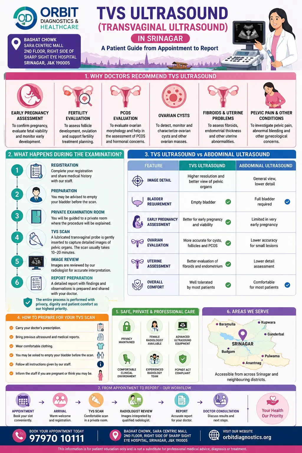
TVS stands for Transvaginal Ultrasound. It is an ultrasound examination that uses a specially designed ultrasound probe to obtain detailed images of the uterus, ovaries, cervix and surrounding pelvic structures. Because the probe is positioned closer to the organs being examined, TVS often provides clearer and more detailed images than a standard abdominal pelvic ultrasound.
Doctors commonly recommend a TVS ultrasound when more detailed pelvic imaging is required. The examination plays an important role in early pregnancy evaluation, fertility assessment, investigation of pelvic pain, abnormal bleeding, ovarian cysts, fibroids and several other gynecological conditions.
Although many patients feel anxious when a TVS scan is recommended, the procedure is routinely performed worldwide and is considered an important diagnostic tool in modern gynecology and women's healthcare.
A TVS ultrasound may be advised when detailed examination of the pelvic organs is needed. The reason for the examination varies from patient to patient depending on symptoms, medical history and the clinical question being investigated.
To confirm pregnancy, evaluate fetal development, assess viability and investigate symptoms such as pain or bleeding during early pregnancy.
To monitor follicle development, assess ovulation, evaluate ovarian function and support fertility treatment planning.
To examine pelvic organs when patients experience pelvic discomfort, lower abdominal pain or unexplained symptoms.
To help evaluate causes of irregular menstrual bleeding, heavy bleeding or post-menopausal bleeding.
To assess ovarian cysts, fibroids, endometrial abnormalities and other gynecological concerns.
To monitor known conditions or evaluate findings identified during previous examinations.
Our team can assist with appointments, examination information and preparation guidance before your visit.
One of the biggest advantages of a TVS ultrasound is the level of detail it can provide when examining pelvic organs. Depending on the clinical situation, the examination may help doctors assess a wide range of gynecological and early pregnancy-related conditions.
Assessment of gestational sac, yolk sac, fetal pole and early pregnancy development.
Evaluation of simple cysts, complex cysts and other ovarian abnormalities.
Assessment of uterine fibroids including size, location and relationship to surrounding structures.
Evaluation of ovarian appearance when polycystic ovarian syndrome is suspected.
Measurement of endometrial thickness and evaluation of abnormalities affecting the uterine lining.
Monitoring follicle growth during fertility assessment and ovulation tracking.
Investigation of possible pelvic causes contributing to pain and discomfort.
Assessment when pregnancy develops outside the uterus and urgent evaluation is required.
Evaluation of possible gynecological causes associated with irregular or heavy bleeding.
Many patients wonder why their doctor recommends a TVS ultrasound instead of a routine abdominal scan. While both examinations use ultrasound technology, they serve different purposes and provide different levels of detail.
| Feature | TVS Ultrasound | Abdominal Ultrasound |
|---|---|---|
| Probe Position | Internal | External |
| Image Detail | Usually higher for pelvic organs | General overview of pelvic structures |
| Bladder Requirement | Usually empty bladder | Often requires a full bladder |
| Early Pregnancy Evaluation | Often preferred for detailed assessment | May have limitations in very early pregnancy |
| Ovarian Assessment | Provides detailed visualization | May provide less detail |
Many women feel nervous before their first TVS ultrasound because they do not know what to expect. Understanding the process beforehand often helps reduce anxiety and makes the experience more comfortable.
Upon arrival, registration details and examination requirements are reviewed. Previous reports may also be checked if available.
The procedure is explained and any questions can be discussed before the examination begins.
The examination is performed in a private clinical environment designed to maintain dignity and confidentiality.
A specially designed ultrasound probe covered with a sterile protective sheath is used to obtain detailed pelvic images.
Images are carefully evaluated and documented for reporting.
The findings are reviewed and the report is prepared according to clinic workflow.
A TVS ultrasound is a personal medical examination, and many patients understandably have concerns regarding privacy. At Orbit Diagnostics & Healthcare, every effort is made to provide a respectful and professional environment where patients feel comfortable discussing sensitive healthcare concerns.
The examination is performed in a private setting with appropriate clinical protocols. Patient confidentiality is respected throughout the process, and examination findings are handled through established reporting procedures.
If you have concerns regarding the examination, preparation requirements or privacy-related questions, you are encouraged to discuss them with the clinic team before your appointment.

MBBS, MD (Radiodiagnosis)
Dr. Ujala Gulzar is a Consultant Radiologist with expertise in diagnostic imaging, ultrasound examinations, Doppler studies, pregnancy imaging and gynecological ultrasound. She completed her MD in Radiodiagnosis from Sher-i-Kashmir Institute of Medical Sciences (SKIMS), Srinagar and was awarded a Gold Medal for academic excellence.
She is currently associated with Paras Health, Srinagar and also serves as a Visiting Consultant Radiologist at Orbit Diagnostics & Healthcare.
This is one of the most common questions women ask after their doctor recommends a TVS ultrasound. In most cases, the examination is not described as painful, although individual experiences can vary.
Most women report feeling mild pressure rather than pain during the examination. The ultrasound probe used for TVS imaging is specially designed for this purpose and is generally inserted gently while the examination is being performed.
Factors such as pelvic tenderness, active pain, anxiety, infection, recent surgery or certain medical conditions may influence how comfortable the examination feels. If you experience discomfort during the scan, it is important to communicate with the healthcare professional performing the examination.
For many patients, the anticipation of the examination is often more stressful than the procedure itself. Understanding the purpose of the scan and knowing what to expect beforehand can help reduce anxiety.
Patients are often concerned when a TVS ultrasound is recommended during early pregnancy. This concern is understandable, especially when the pregnancy is newly confirmed or when symptoms such as pain or bleeding are present.
TVS ultrasound uses sound waves to create images and does not involve ionising radiation. When performed for appropriate medical reasons, it is widely used in early pregnancy assessment throughout the world.
Doctors may recommend a TVS scan during early pregnancy because it can provide detailed information regarding the gestational sac, fetal pole, yolk sac and fetal heartbeat at a stage when these structures may not yet be clearly visible on an abdominal ultrasound.
The examination should always be interpreted in the context of the patient's medical history, symptoms and the advice of the treating healthcare professional.
Preparation requirements can vary depending on the reason for the examination. The clinic team may provide specific instructions when your appointment is scheduled.
Unlike many abdominal pelvic ultrasounds, a TVS ultrasound often does not require a full bladder. However, preparation instructions may differ depending on the clinical indication, so patients should follow the guidance provided for their individual appointment.
If the examination has been advised by a doctor, bringing the prescription can help provide useful clinical information.
Earlier ultrasound reports, laboratory results or relevant medical records may help with comparison and follow-up evaluation.
Patients may be requested to provide identification and other information required for registration and regulatory compliance.
Referral letters and treatment summaries can provide valuable context for the examination.
Many patients receive their report and immediately start searching online for unfamiliar medical terms. While final interpretation should come from your doctor, understanding a few common terms may help you better follow the discussion.
The report may describe the size, shape and appearance of the uterus.
This refers to the lining of the uterus and may be measured depending on the reason for the examination.
The appearance, size and any visible cysts or follicles may be described.
Follicle measurements may be included when fertility monitoring is being performed.
If present, fibroids may be described according to their size and location.
In pregnancy-related examinations, findings such as gestational sac, fetal pole and cardiac activity may be reported when appropriate.
It is important to remember that a report is only one part of the diagnostic process. Your treating doctor will interpret the findings together with your symptoms, medical history, examination findings and any additional investigations.
One of the most common questions patients ask before booking an appointment is the expected cost of a TVS ultrasound. The exact charges can vary depending on the clinical indication, reporting requirements and whether additional imaging evaluation is required.
Orbit Diagnostics & Healthcare aims to provide transparent information regarding diagnostic services. For the most accurate and current pricing information, patients are encouraged to contact the clinic directly.
Our team can also explain appointment availability, preparation requirements and report timelines before scheduling your visit.
Many women feel anxious when a TVS ultrasound is recommended, especially if it is their first experience with the examination. Questions about privacy, dignity and comfort are completely understandable.
At Orbit Diagnostics & Healthcare, patient comfort is treated as an important part of the examination process. Every effort is made to provide a respectful environment where patients can discuss sensitive healthcare concerns comfortably and confidently.
The examination is performed in a private clinical setting. Patients are encouraged to ask questions before the scan and discuss any concerns they may have regarding the procedure.
Our goal is not only to provide diagnostic imaging but also to ensure that patients feel informed, respected and supported throughout their visit.
Orbit Diagnostics & Healthcare strictly follows the provisions of the Pre-Conception and Pre-Natal Diagnostic Techniques (PCPNDT) Act and applicable regulations governing ultrasound practice in India.
Pre-natal sex determination and disclosure of fetal sex are prohibited by law and are not performed or supported at our facility.
Patients may be required to provide documentation, identification and information necessary for regulatory compliance before certain examinations.
Patients regularly visit Orbit Diagnostics & Healthcare from different areas of Srinagar and neighbouring districts for ultrasound examinations, Doppler studies and diagnostic imaging services.
The clinic is conveniently located at Baghat Chowk, Barzulla and is accessible from Rawalpora, Hyderpora, Peer Bagh, Chanapora, Natipora, Rambagh, Sanat Nagar, Nowgam, Bemina, Tengpora, Humhama, Rangreth and surrounding areas.
Patients also travel from Budgam, Ganderbal, Pulwama, Anantnag, Kulgam, Shopian, Baramulla, Bandipora and Kupwara when seeking diagnostic imaging services in Srinagar.
Ultrasound examinations are reviewed by qualified radiology professionals with expertise in diagnostic imaging.
Support for pregnancy imaging, gynecological ultrasound and fertility-related evaluation.
Located at Baghat Chowk, Srinagar with access from multiple nearby areas.
Respectful communication and privacy-focused examination workflow.
Our team can assist patients with scheduling, preparation guidance and service information.
Dedicated focus on ultrasound and diagnostic imaging services.
Whether you have been referred by your doctor, require fertility monitoring or need pregnancy-related imaging, our team can help you understand the next steps and arrange an appointment.
Patients often have questions about preparation, safety, privacy, pregnancy, fertility and what to expect during a TVS ultrasound examination. The answers below address many of the common concerns we hear from women before their appointment.
Medical content published by Orbit Diagnostics & Healthcare is reviewed periodically to help ensure accuracy, relevance and alignment with accepted diagnostic imaging practices.
Lead Medical Reviewer
Dr. Ujala Gulzar
MBBS, MD
Note: Content is intended for educational purposes and does not replace professional medical advice, diagnosis or treatment.
Dr. Ujala Gulzar
MBBS, MD (Radiodiagnosis)
Consultant Radiologist
Health information presented on this page has been prepared for educational purposes and may be informed by established radiology references, diagnostic imaging literature, clinical practice guidance and accepted ultrasound principles.
This page is intended to support patient understanding and should not replace professional medical advice, diagnosis or treatment.
Healthcare decisions often involve more than choosing the nearest facility. When a detailed pelvic ultrasound examination is required, many patients prefer a centre where they can access experienced radiology support, professional reporting and clear communication throughout the process.
Orbit Diagnostics & Healthcare welcomes patients from different parts of Srinagar as well as neighbouring districts across Kashmir. While many visitors come from areas such as Baghat Chowk, Barzulla, Rawalpora, Hyderpora, Chanapora, Natipora and Rambagh, patients also travel from Budgam, Ganderbal and Pulwama for ultrasound and diagnostic imaging services.
Women referred for fertility assessment, early pregnancy evaluation, pelvic pain investigation or follow-up gynecological imaging may also visit from districts including Anantnag, Baramulla and Kupwara. Some patients are referred by their treating doctors, while others choose the clinic based on location convenience, appointment availability or previous experience with diagnostic services.
Because TVS ultrasound examinations are often performed for sensitive and personal healthcare concerns, many patients value having access to a professional clinical environment where privacy, respectful communication and accurate diagnostic reporting remain important priorities.
Our location at Baghat Chowk, Srinagar helps make the clinic accessible for patients travelling from different parts of the Kashmir Valley. The clinic is situated near major connecting routes, allowing patients from both Srinagar city and surrounding districts to reach the facility without unnecessary difficulty.