Diagnostic Imaging Guide

When Is Ultrasound Used Instead Of A CT Scan?

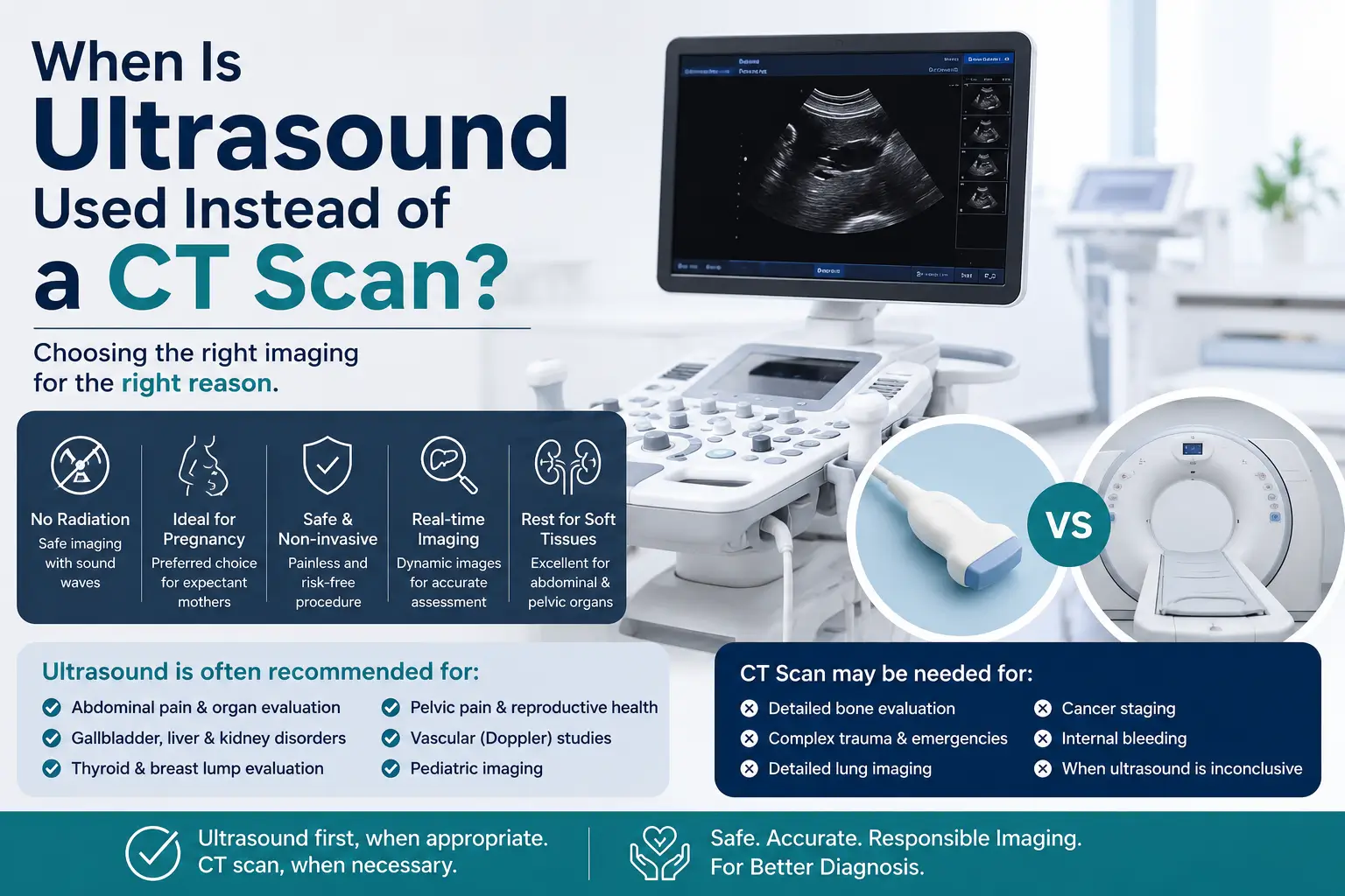
Patients are often surprised when a doctor requests an ultrasound instead of a CT scan. Many assume that CT technology is always more advanced and therefore automatically better. In reality, medical imaging does not work that way. The best imaging test depends on the organ being examined, the patient's age, the clinical question being asked, and whether exposure to radiation can be avoided. In many situations, ultrasound provides the information a clinician needs without requiring ionizing radiation or contrast injections. Understanding why ultrasound is chosen in certain situations can help patients feel more informed before their examination.

Why The Choice Of Imaging Matters

Modern healthcare has access to several imaging technologies. Ultrasound, CT, MRI and X-ray each contribute unique information. Rather than asking which test is superior, doctors usually ask a different question: which examination is most appropriate for the problem being investigated? A person with abdominal pain may receive an ultrasound. Another patient with severe trauma may require an urgent CT scan. A pregnant woman may undergo multiple ultrasound examinations throughout pregnancy without ever needing CT imaging. The decision is guided by clinical usefulness rather than technology alone. For many common medical situations, ultrasound is often selected as the first imaging investigation because it is widely available, produces real-time images and avoids radiation exposure.

Ultrasound versus CT scan comparison for pregnancy, abdominal and diagnostic imaging
A visual comparison showing situations where ultrasound may be preferred over a CT scan, including pregnancy imaging, abdominal evaluation, thyroid assessment and Doppler studies.

What Is Ultrasound?

Ultrasound uses high-frequency sound waves to create images of structures inside the body. A handheld probe sends sound waves into tissues and receives returning echoes. A computer converts those echoes into images that can be viewed instantly. One of the major advantages of ultrasound is that the examination occurs in real time. Organs can be observed while moving, blood flow can be assessed using Doppler technology, and patients can often interact with the examiner during the study. Ultrasound is commonly used for pregnancy assessment, abdominal imaging, thyroid evaluation, vascular studies, soft tissue examination and many other clinical applications. A question arises in many patients: Is Ultrasound Safe During Pregnancy?

What Is A CT Scan?

Computed Tomography, commonly known as CT scanning, combines multiple X-ray images acquired from different angles to create detailed cross-sectional views of the body. CT technology is particularly valuable when physicians need a broad overview of internal anatomy, especially in emergency situations. The examination can rapidly evaluate the brain, chest, abdomen, pelvis and bones. Although CT is an extremely important diagnostic tool, it uses ionizing radiation. For this reason, healthcare professionals generally avoid unnecessary CT examinations when another imaging technique can answer the same clinical question.

When Doctors Often Choose Ultrasound First

There are many situations where ultrasound may be the preferred initial investigation. The reasons vary from one clinical scenario to another. In some cases the target organ is simply easier to evaluate with ultrasound. In others, avoiding radiation exposure becomes an important consideration. The following sections examine some of the most common situations where ultrasound is frequently selected before CT imaging.

Pregnancy Imaging

Pregnancy is one of the clearest examples of why ultrasound is frequently preferred over CT scanning. Obstetric ultrasound has become a routine part of prenatal care because it allows healthcare professionals to evaluate fetal growth, fetal position, placental location and pregnancy development without exposing the mother or fetus to ionizing radiation. Throughout pregnancy, ultrasound can be repeated whenever clinically required. Doctors may request first-trimester scans, anomaly scans, growth scans and Doppler examinations depending on the circumstances. Because ultrasound provides detailed information about pregnancy while avoiding radiation exposure, it remains the primary imaging tool in obstetric practice worldwide.

Gallbladder Disease And Gallstones

When patients experience pain in the upper right portion of the abdomen, doctors often suspect gallbladder disease. In such situations, ultrasound is frequently the first imaging examination requested. Gallstones are often visible on ultrasound because stones create characteristic acoustic shadows that can be recognized during the examination. Ultrasound also allows visualization of gallbladder wall thickening, fluid around the gallbladder and other findings that may help clinicians evaluate gallbladder inflammation. In many routine gallbladder investigations, ultrasound provides the information required without the need for CT imaging.

Liver Evaluation

The liver is another organ commonly assessed using ultrasound. Physicians may request liver ultrasound when investigating abnormal liver function tests, abdominal discomfort, fatty liver changes or enlargement of the liver. Ultrasound can demonstrate differences in liver texture, identify cysts, detect certain masses and evaluate blood flow using Doppler techniques. Modern ultrasound systems can also support specialized assessments in selected clinical settings. Although CT and MRI remain important for certain complex liver conditions, ultrasound often serves as the first-line examination because it is accessible, non-invasive and radiation free.

Kidney And Urinary Tract Assessment

Ultrasound is frequently used when evaluating the kidneys, urinary bladder and urinary tract. Patients presenting with flank pain, urinary symptoms or suspected obstruction may undergo ultrasound as an initial investigation. The examination can help identify kidney enlargement, urinary retention, bladder abnormalities and hydronephrosis, a condition where urine accumulates within the kidney because of impaired drainage. Although CT may be required in selected situations, particularly when stone detection becomes a priority, ultrasound is often the first step because it provides valuable information without radiation exposure.

Thyroid Imaging

The thyroid gland is located close to the skin surface, making it particularly suitable for ultrasound examination. As a result, ultrasound has become the primary imaging method for evaluating many thyroid-related concerns. Doctors commonly request thyroid ultrasound when a patient develops a neck swelling, thyroid enlargement or a thyroid nodule. The examination can help characterize the size, shape and internal appearance of thyroid abnormalities. Because the thyroid is easily accessible to sound waves, CT scanning is usually not the first imaging choice for routine thyroid assessment.

Blood Vessel Evaluation With Doppler Ultrasound

One of the unique advantages of ultrasound is the ability to assess blood flow in real time through Doppler technology. Color Doppler and spectral Doppler examinations can help evaluate arteries and veins throughout the body. These studies are commonly used when investigating leg swelling, suspected venous disorders, carotid artery disease and other vascular conditions. Unlike CT angiography, Doppler ultrasound does not necessarily require contrast administration and avoids radiation exposure. For many vascular investigations, it serves as an important first-line diagnostic tool.

Children Often Benefit From Ultrasound First

Medical imaging decisions in children frequently place greater emphasis on minimizing radiation exposure. Because growing tissues may be more sensitive to radiation, healthcare providers often prefer ultrasound whenever it can answer the clinical question. Pediatric abdominal pain, suspected appendiceal abnormalities, urinary tract concerns and various soft tissue conditions may initially be investigated with ultrasound. The goal is not simply to avoid CT scanning, but rather to use the most appropriate examination while limiting unnecessary radiation whenever possible.

Soft Tissue Lumps And Surface Abnormalities

Patients occasionally notice lumps beneath the skin and wonder what type of imaging might be required. In many cases, ultrasound serves as the first examination because it is particularly effective for evaluating superficial structures. The examination can help determine whether a lump appears solid, fluid-filled or mixed in nature. Ultrasound may also assist clinicians in understanding the relationship between the abnormality and nearby tissues. Because these structures are often located close to the skin surface, ultrasound frequently provides excellent visualization without requiring CT imaging.

Why Doctors Prefer To Avoid Radiation When Appropriate

CT scanning remains one of the most valuable advances in modern diagnostic medicine. However, because CT uses ionizing radiation, healthcare providers generally follow the principle of using the lowest reasonable radiation exposure necessary to answer a medical question. When ultrasound can provide the required information, choosing ultrasound may help avoid radiation altogether. This consideration becomes particularly important in pregnancy, childhood imaging and situations where repeat examinations may be necessary over time. The decision is not based on convenience. Instead, it reflects a balance between diagnostic accuracy, patient safety and clinical necessity.

Situations Where CT Scan May Still Be Preferred

Despite the many advantages of ultrasound, there are numerous circumstances where CT remains the preferred investigation. Emergency trauma assessment, evaluation of certain lung conditions, complex abdominal emergencies, detailed bone imaging and many cancer-related investigations often require CT scanning because of its ability to rapidly produce comprehensive cross-sectional images. For this reason, ultrasound and CT should not be viewed as competing technologies. They are complementary diagnostic tools, each serving different clinical purposes. The most appropriate examination depends on the individual patient, symptoms, medical history and the diagnostic information needed by the treating physician.

Ultrasound Vs CT Scan: Quick Comparison

Feature Ultrasound CT Scan
Technology Sound Waves X-Ray Based Imaging
Radiation Exposure No Yes
Pregnancy Imaging Commonly Used Usually Avoided Unless Necessary
Real-Time Imaging Yes Limited
Blood Flow Assessment Color Doppler Available May Require CT Angiography
Gallbladder Evaluation Frequently First Choice Selected Situations
Thyroid Examination Frequently First Choice Less Common
Emergency Trauma Limited Role Frequently Preferred

Frequently Asked Questions

Is Ultrasound Safer Than A CT Scan?

Ultrasound does not use ionizing radiation. However, the most appropriate imaging examination depends on the medical condition being investigated. Doctors select the examination that can best answer the clinical question.

Why Would A Doctor Request Ultrasound First?

Ultrasound may provide sufficient diagnostic information while avoiding radiation exposure. It is frequently used for pregnancy, gallbladder, thyroid, vascular and many abdominal examinations.

Can Ultrasound Replace CT Scans Completely?

No. CT scanning remains essential for many medical situations including trauma assessment, complex emergencies, certain cancers and detailed cross-sectional evaluation of internal structures.

Can Blood Flow Be Seen On Ultrasound?

Yes. Doppler ultrasound technology can evaluate blood flow within arteries and veins and is widely used in vascular imaging.

Which Test Is Better?

Neither test is universally better. Ultrasound and CT serve different purposes. The most suitable examination depends on the symptoms, body region and information needed by the treating clinician.

Final Thoughts

Many patients assume that newer or more complex imaging automatically produces better results. In practice, medical imaging is about choosing the right tool for the right question. Ultrasound remains one of the most widely used diagnostic examinations in modern medicine because it provides real-time imaging, avoids radiation exposure and performs exceptionally well in many clinical situations. Pregnancy assessment, gallbladder disease, thyroid imaging, vascular studies and numerous abdominal examinations often begin with ultrasound for these reasons. CT scanning continues to play a vital role in healthcare and remains indispensable for many conditions. Rather than competing with each other, ultrasound and CT work together as complementary diagnostic technologies that help clinicians make informed decisions. Understanding why a physician selects one examination over another can help patients approach imaging investigations with greater confidence and clarity.

Medical content across Orbit Diagnostics & Healthcare is reviewed and maintained by qualified healthcare professionals and diagnostic imaging specialists where applicable.

Lead Medical Reviewer:
Dr. Nidha Nazir
Consultant Radiologist
Orbit Diagnostics & Healthcare

Learn More About Our Team